A system known as the digestive system has been developed in vertebrates to perform the functions of food swallowing, digestion, absorption and assimilation and excretion of indigestible parts. Most of the food changes and becomes simple and soluble so that it can be easily absorbed by the blood and lymph. Such mechanical and chemical breakdown or change in food is called digestion.
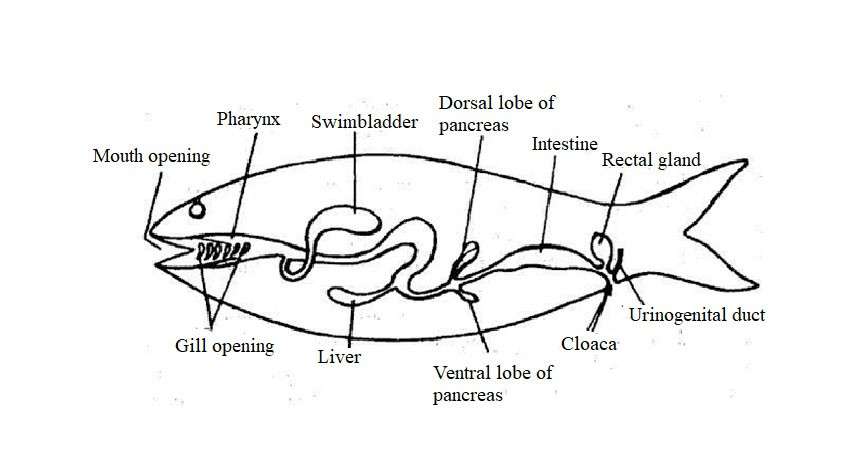
The digestive system of fish consists of the digestive tract and various associate organs such as the tongue, teeth, oral glands, pancreas, liver and gallbladder. The digestive tract consists of the anterior, middle and posterior parts. Each part is then divided or converted into several pieces. The anterior part is transformed into the mouth, mouth cavity, pharynx and stomach. The middle part changes to form the small intestine and the posterior part forms the large intestine and the anus.
Fig. Showing Outline diagram of digestive system of Fish
Digestive Tract
The digestive tract of fish consists of different parts. Modification of the digestive tract occurs due to differences in the diet and diet of fish. Adaptation of the fish’s digestive tract and its various parts are described below:
Lips: Usually the mouth is surrounded by lips. Food-sucking fish such as Sturgeon ( Acipenser), Labeo, Cirrhinus and Puntius have fleshy lips. Many fish, including the Sisurid catfish (Glyptosternum) and the South American armored fish (Loricaridae), they have transformed their beaks to form a hold fast organ-like sucking disc.
Mouth and Jaws: The mouth of the fish is opening type. Different species of fish undergo mutations based on the position of the mouth, shape, size, dietary characteristics, food intake and eating habits. Jawed fish have jaws around their mouths. Fish jaws were first developed in Acanthodian. The transformation of the actual jaw occurs with the change from microscopic to large-scale food intake.
Differences of mouth are seen in different fish. It is usually marginal, but in many groups, such as Chondrichthyes and Siluroids, it exists on the ventral side. Mouth position is located at the top of the mouth in predator fish like characinoids and some hemorrhamphids (Hyporhampus limbatus). The mouth is located on the ventral side in the bottom-dwelling fish, on the other hand, the surface feeder fish have mouth on the dorsal side but the column feeder fish have terminal mouth.
Different fish have different shaped mouth. The jaws of the mouths of grazing and sucking fish have changed to form elongated whistle structure. Fistularia villosa, pipe fish (Syngnathus), Xenentodon cancila and butterfly fish (Forcipiger longirostris) have long beaked mouths. The long beaks of Fistularia and Syngnathus suck food like syringes and the toothed lips of Forcipiger and Xenentodon help in finding food.
In the Hyporhampus, only the lower jaw forms long bony lips. The upper jaw is small and it forms a triangular stretched part. These types of fish are surface feeder and their beaks help to balance the body and collect the food using the dorsal fins.
Various species of Nandus and Channa have more expandable mouth gape while Anabas has narrowly expandable mouth gape.
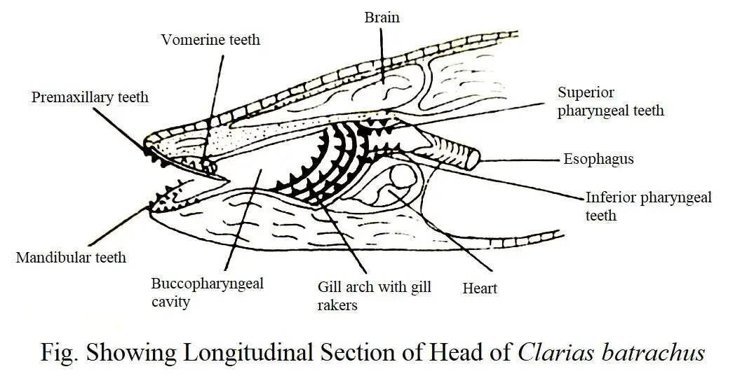
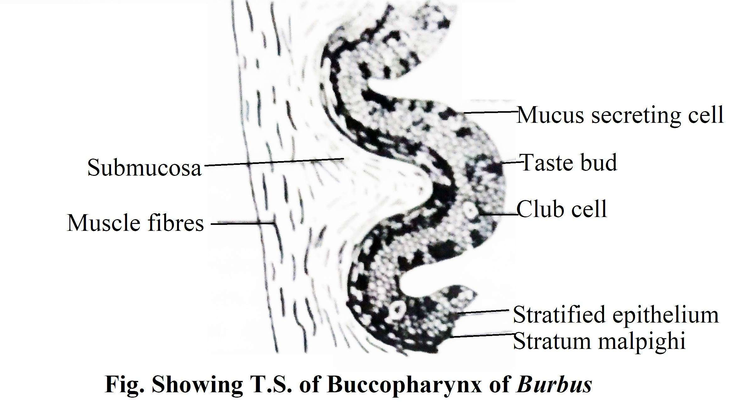
Buccal cavity and Pharynx: The mouth cavity and the pharynx cannot be clearly distinguished. There are a significant number of gill opening on each side of the pharyngeal wall. These are exposed to the gills through the gill rakers and help in food intake. The pharynx is a larger structure for respiration than food digestion. The mouth cavity holds the teeth and the tongue.
Teeth: Teeth of the vertebrates can be divided into two main types, epidermal and dermal. Epidermal teeth originate from the stratum corneum of the epidermis, and such teeth are confined to the oral funnel and tongue of Agnatha. Dermal teeth are derived from dermis. The placoid scales of most mammals and many sharks are constantly transformed into teeth and placed adjacent to the jaw edges.
Like placoid scales, these teeth of fish have a cone-shaped hollow dentin coating with pulp cavity. These are produced continuously in the form of special denticles or placoid scales along the inner edges of the jaw. The new tooth is generated behind the old tooth and pushes the old tooth forward. As a result, when the old teeth are eroded, new teeth are produced from the back. Different fish vary in number of teeth, width, degree of stability, shape and type of tooth attachment.
The buccopharynx of fish has two functions namely:
(1) respiration and
(2) catching food and reaching the esophagus.
In chondrichthyes, the teeth are scattered on the roof of the mouth or they are attached to the cartilaginous jaw with the help of connective tissue. These teeth are called acrodont. These teeth look similar in size and shape (homadont) and may be replaced by posterior teeth (polyfiodont).
Osteichthyes teeth can be divided into three groups based on their location in the oral cavity. The premaxillary and maxillary bones carry the jaw teeth upwards with the dentin below. Vomar bones carry the oral teeth. The palatine and ectopterygoid bones carry the roof teeth and the tongue carry the floor teeth of the mouth. There are three types of pharyngeal teeth that erupt from different sources such as the lower component of the last gill arch, gill rakers or dermal bone.
There are different types of teeth of fish based on eating habits. Teeth are pointed, curved, cutting type, crushing type or venom-like teeth of snake. In some cases, in particularly the Dipnoins, they have budding teeth which combine to form plate-shaped composite teeth that are used to crush food.
Carnivorous and predatory fish such as Wallago attu, Mystus seengala, M. aor, Channa marulius, C. punctatus, C. gachua, Chitala chitala (=Notopterus chitala), Harpodon nehereus, Muraenesox talobon have strong teeth in different parts of the buccopharynx. Even their gill rakers are look like teeth. Their buccopharynx is not precisely divided into the buccal cavity and pharynx.
The anterior edge of the gill is called the buccal cavity. Premaxilla, maxilla, vomer, palatine and dentery contain teeth. Some fish such as Chitala chitala (= Notopterus chitala) and N. notopterus have teeth on their tongues. The pharyngeal tooth rests on the gill arch and is known as the superior and inferior pharyngeal teeth. Teeth are of villiform, incisiform, canine-like and molariform types. These teeth are sharp, curved and pointed. They are not used in crushing food. The front teeth of the mouth cavity prevent the prey from escaping. Pharyngeal teeth help to prevent discharge the prey. Moreover, it also acts as a crushing organ.
In herbivorous fish such as Labeo rohita, L. gonius, L. calbasu and some omnivorous fish, the buccopharynx are divided into anterior respiratory tract and posterior narrow region (which is used as a chewing organ). These fish do not have teeth in their jaws and palate, but they have well-developed subpharyngeal teeth. The fifth cerotobranchial carries these teeth. It crushes the prey by acting against the dorsal hard calcareous pad through the besi-occipital bone. Intermediate conditions are seen in omnivorous fish.
Some species such as Eutropichthyes vacha, Clarias batrachus have jaw and palate teeth. However, it is less developed than carnivores. Some fish such as Tor tor, Puntius sarana, Catla catla do not have jaw and palate teeth. However, the mouth cavity is like a herbivorous fish. Plankton eaters such as Tenualosa ilisha, Gadusia chapra have no teeth in the pharynx. However, some fish such as Atherina forskali, Ilisha filigera have small teeth in their jaws.
Tongue: The tongue originates as a fold from the floor of the mouth cavity. It has no muscles but extends inward with the help of a hyoid arch. On this structure, tiny papillae, sensory receptors and teeth exist.
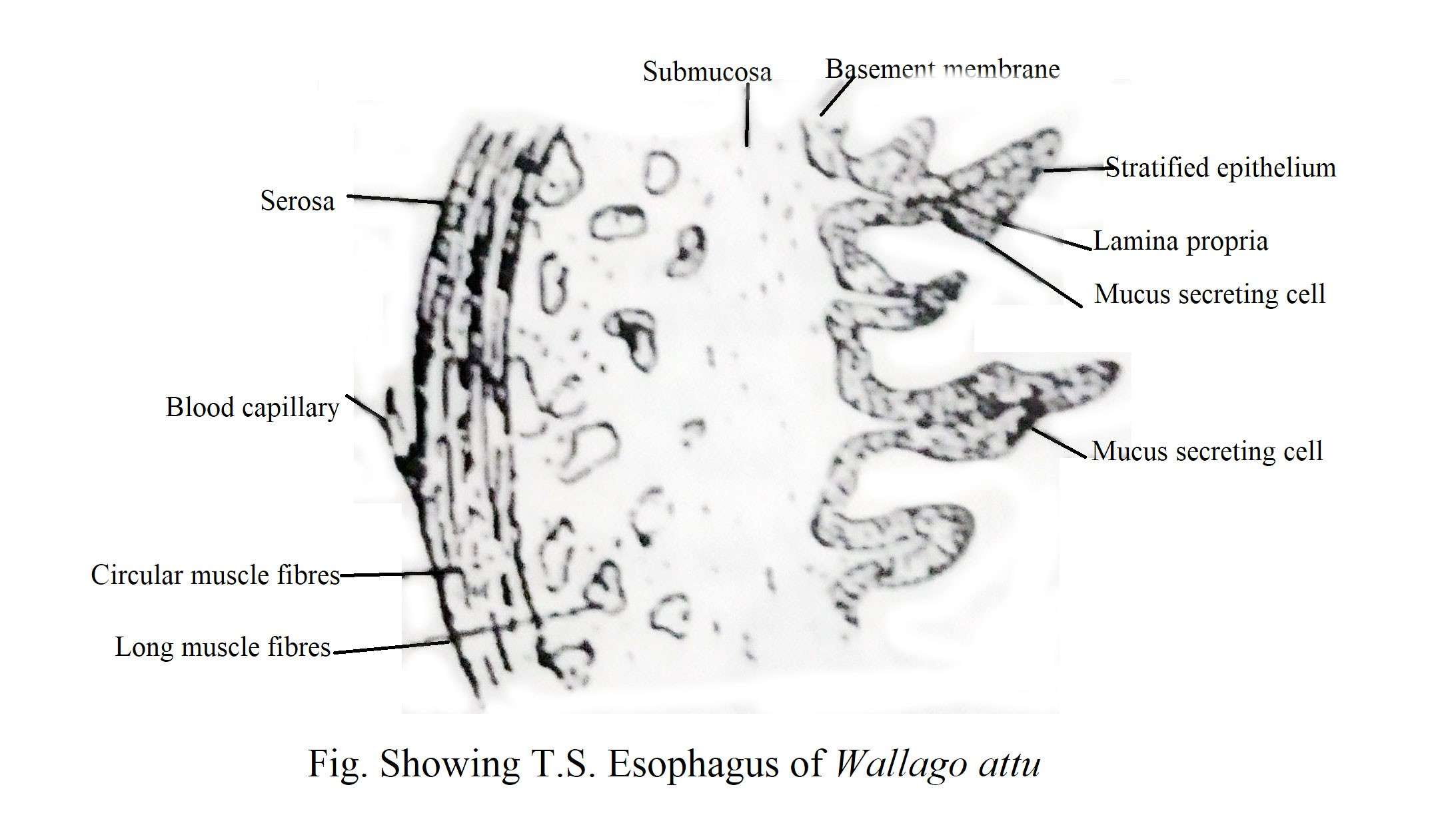
Esophagus: The pharynx is exposed to the esophagus at the back and later to the stomach in most cases. In most cases, the pharynx, esophagus, and stomach cannot be clearly distinguished without histological structure. The esophagus is usually swollen due to its longitudinal folds and its mucous membrane is made up of squamous cells.
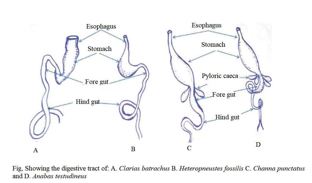
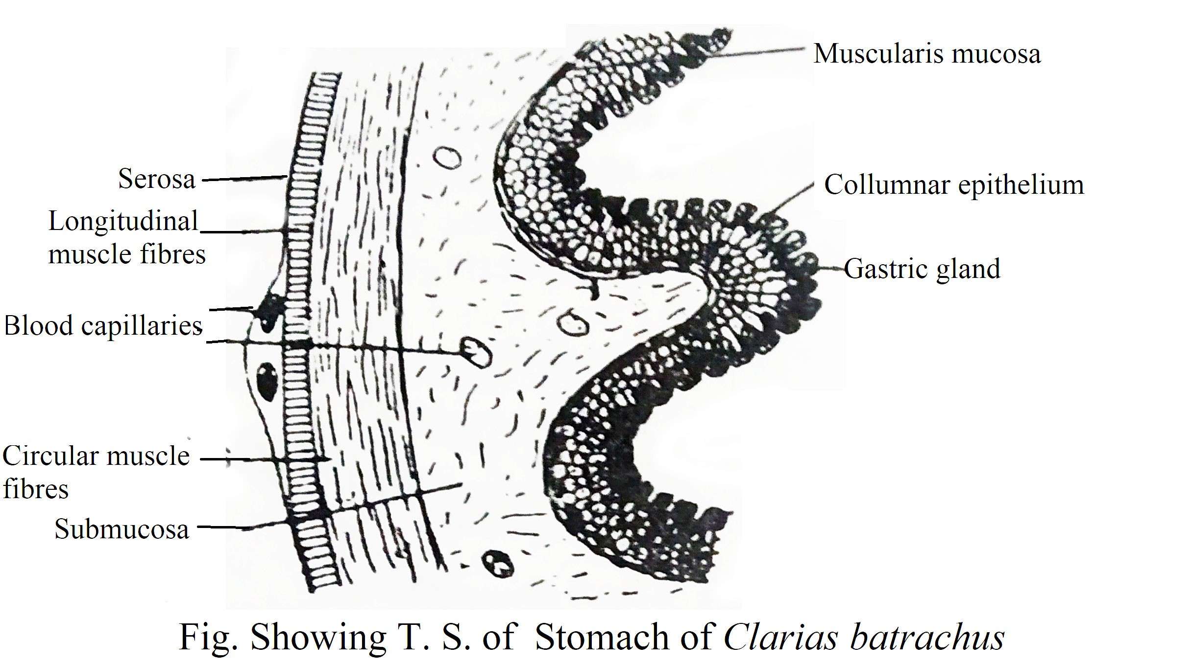
Stomach: The stomach is basically a structure that is engaged in storing and crushing food. When there is a gastric gland in the inner wall of the stomach, it is called true stomach. Externally, the stomach cannot be separated from the esophagus. They can be separated from the folds of the mucosa. The folds of the esophageal mucosa are thin while in the stomach, the mucosal fold is thick with wavy edge.
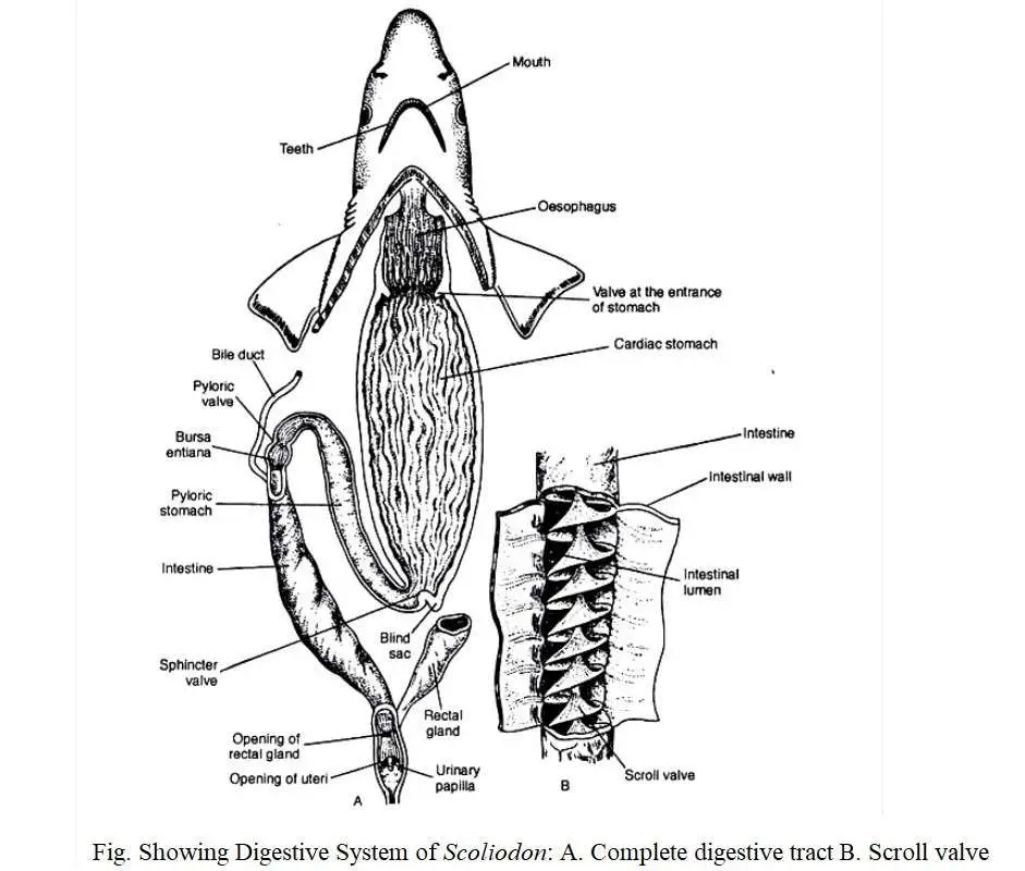
The stomachs come in different shapes depending on the size of the body cavities of different fish. It is mainly divided into two parts, namely, cardiac stomach-the wide part of the anterior portion of the stomach which is located near the heart and the pyloric stomach-the posterior narrow opening. The pyloric stomach is connected to the mid gut through an opening that is controlled by a valve.
In Elasmobranch, the stomach has a standard ‘J’ shaped structure which is divided into two parts. The upper long and broad part is known as cardiac and the short below part is known as pyloric stomach. Many fish have a circular fold in the mucous membrane at the junction of the cardiac and pyloric stomachs that form the pyloric valve. Some fish (Scoliodon) have a small thick-walled small chamber which is known as bursa entiana at the end of the pyloric stomach. The stomach contains gastric glands that cover the mucous membranes.
The stomach of the teleost comes in different shapes. In some cases, no distinction can be made between the esophagus and the stomach due to its same diameter. In other cases, a round and muscular structure is seen at the end of the esophagus so that there are well-formed gastric glands. The stomach of some other fish is divided into cardiac and pyloric, with closed sacs of different sizes. Fish of the family Cyprinidae and Catostornidae have no true stomach and the digestion process is done into the intestine by pancreatic juice. In the fish of Cobitidae family, there are a large number of fish with and without stomach.
Carnivorous and predatory species such as Wallago attu, Mystus senghala, Channa striatus, Chitala chitala (= Notopterus chitala), Harpodon nehereus, Muraenesox talabon, etc., have sac like with thin walled stomachs. Some species such as Tenualosa ilisha, Mugil, Gadusia chapra have greatly reduced stomachs and its wall has become too thick to form a gizard-like structure for food absorption.
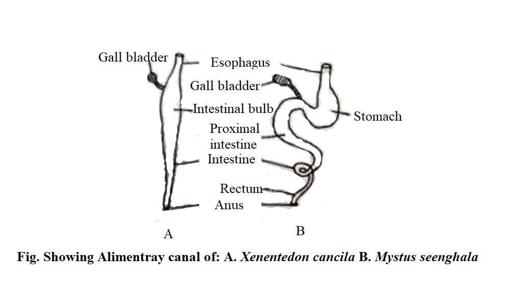
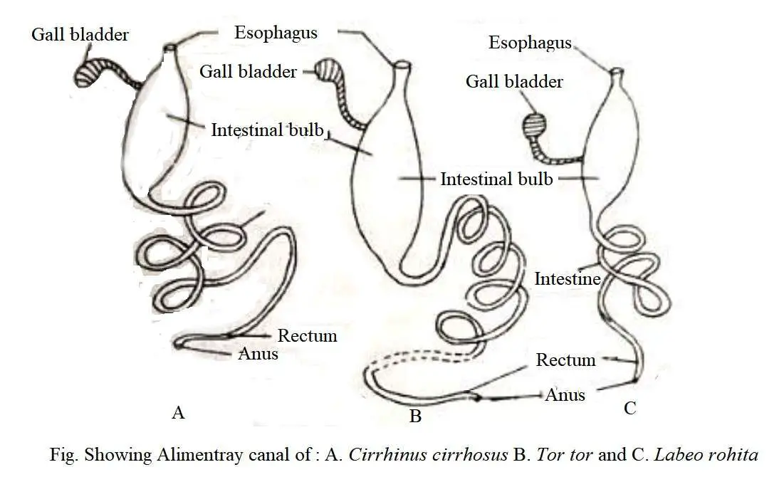
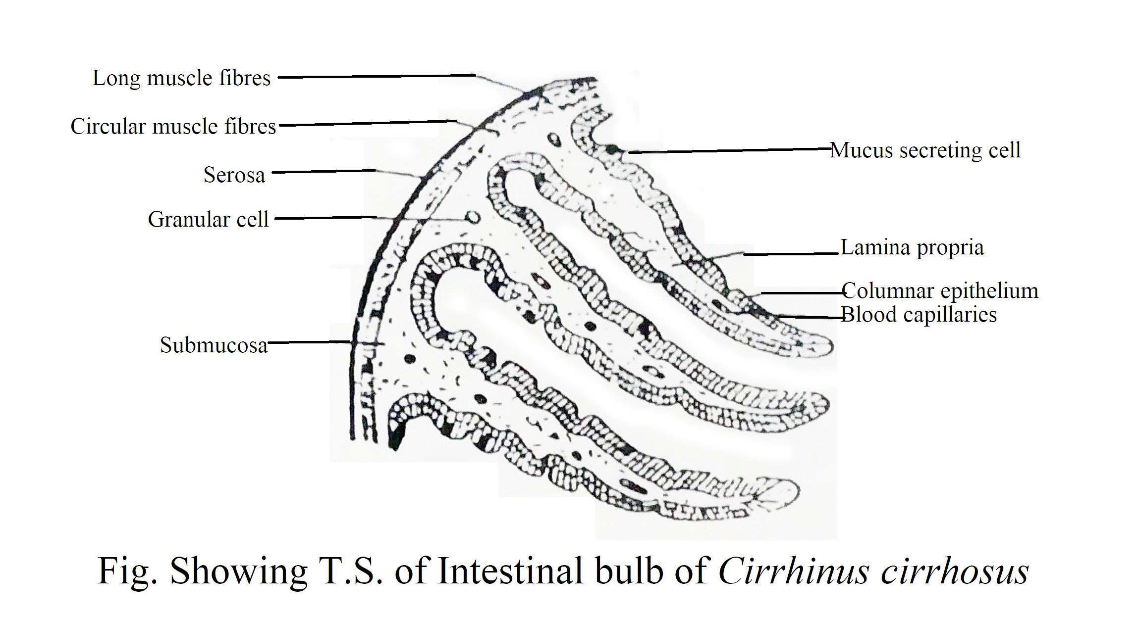
Not all types of fish have true stomachs and some fish do not have stomachs. In Labeo gonius, Cirrhina cirrhosus, Tor tor, Ctla catla, Puntius sophore, etc. the anterior part of the intestine behind the esophagus swells and form a sac-like structure which stores food and is known as intestinal bulb. The presence or absence of true stomach does not depend on the diet of the fish.
Holocephali and Dipnoi also do not have stomach. Apart from Cyprinid, some other fish like Scomberesox, Xenentodon, Hippocampus do not have stomach. The intestinal bulb does not have a gastric gland and its mucosa is closely related to the intestine. Absorptive cells and mucus-secreting cells are present in the mucosal epithelium of the intestinal bulb.
In the case of lung fish, the esophagus and stomach can be separated by a constriction. These fish do not have gastric glands in their stomach, but they are separated from the intestine by a filamentous pyloric valve.
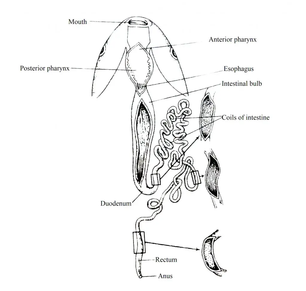
Intestine: The part of the esophagus behind the stomach is called the intestine. It is divided into two main parts. The long and narrow duct of the anterior part is called the small intestine which is located just behind the stomach and here the ducts coming from the liver and pancreas meet together and it is called duodenum. The next part of the small intestine is called the ileum. All of these different types of intestinal tract are usually histologically distinct only as a result of continuous changes in the mucosa level. Only in some group of fish, a fold is present behind the intestine.
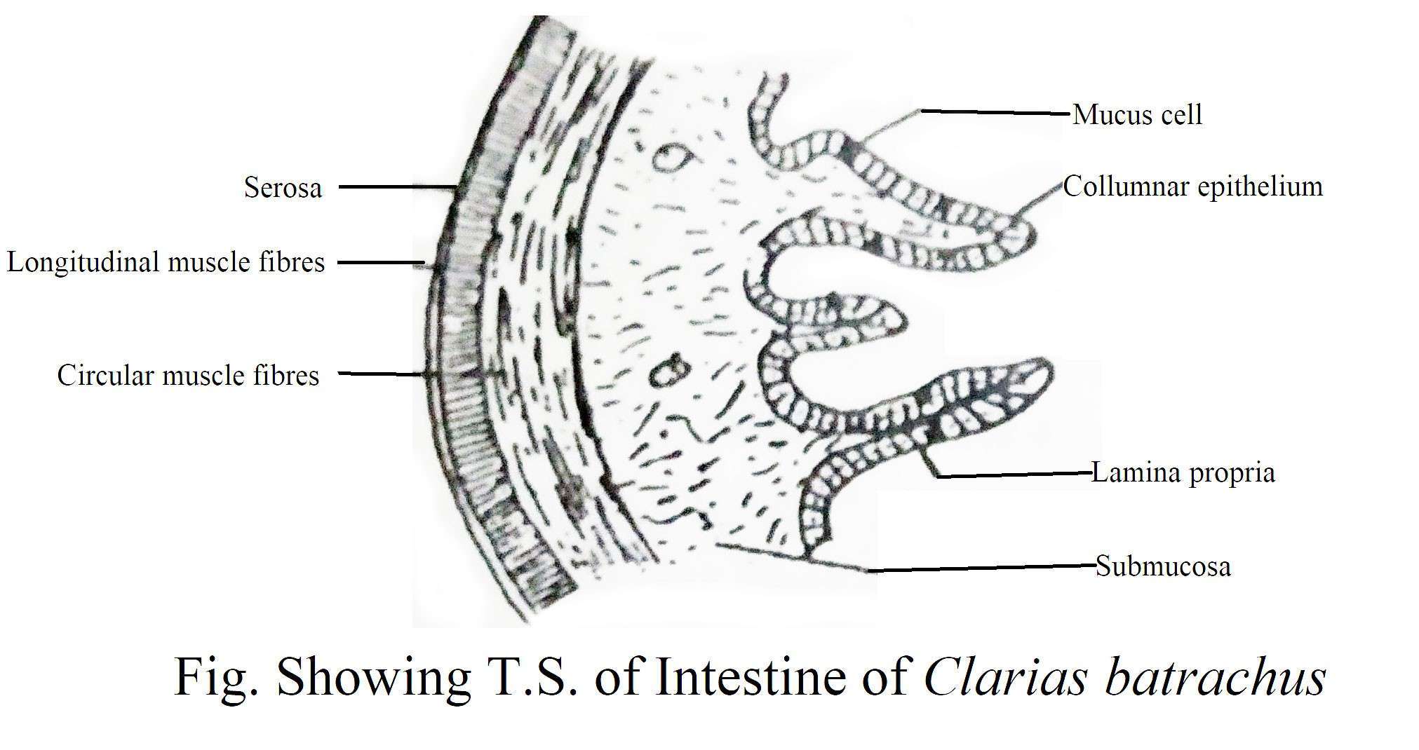
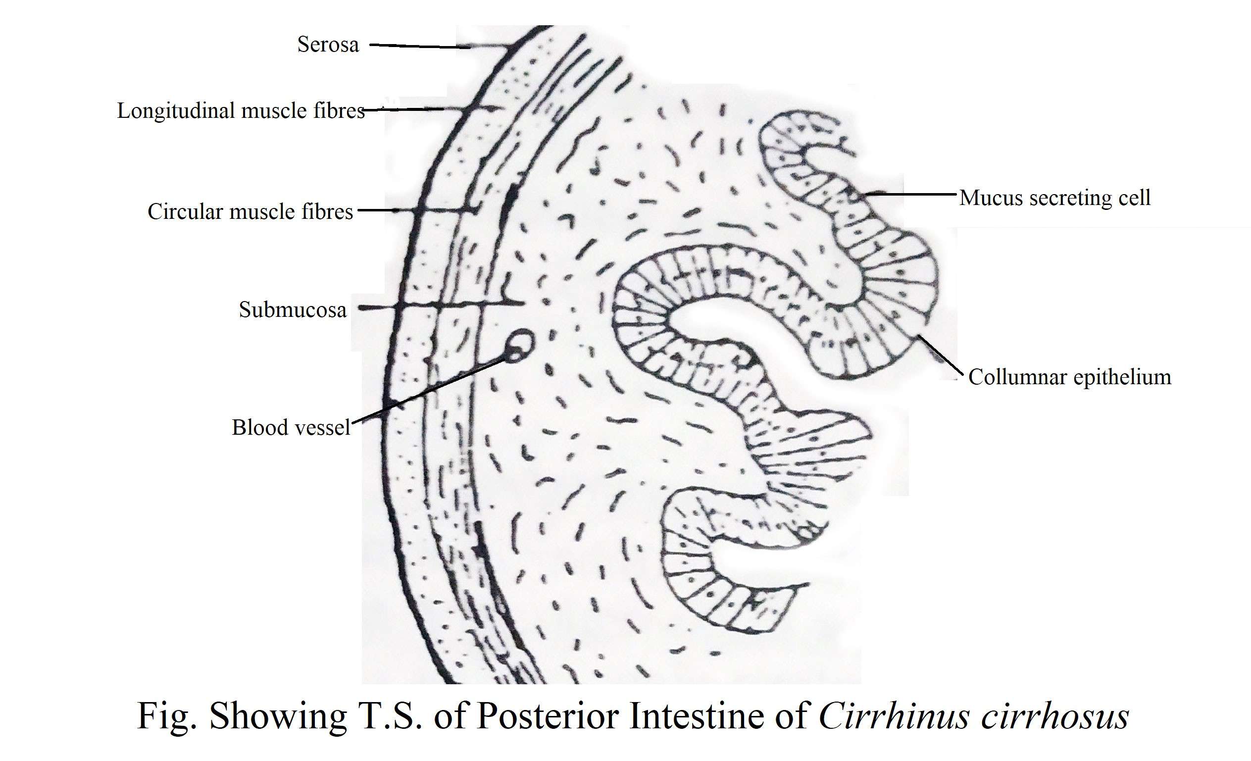
The length of intestine varies in different species of fish. In carnivorous fish, it is usually short and fairly straight. However, in herbivorous species such as Labeo and Cirrhina, the intestines are much longer and more coiled. The intestinal wall contains absorptive and mucus-secreting cells which are stituated in a simple columner epithelium layer. The anterior part of the intestine contains a small number of mucus-secreting cells, rarely in the central region, but the number of these cells increases in the posterior part.
In some species, the mucosal fold is branched. The intestinal mucosa also contains a very small number of glandular cells that are basophilic or acidophilic in nature but their function is unknown. The intestinal muscularis layer is thin and they are composed of outer longitudinal and inner circular muscular layer.Transition condition is found in omnivorous species. According to Das and Moitra (1958), the ratio of intestinal length and body length of most fish is fairly stable. However, some carnivorous fish such as Pterois have a long looped intestine.
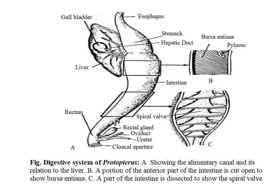
According to Suyehiro (1942), in most omnivorous fish, there is no relationship between length of intestine and food nature. According to Barrington (1956), the relative length of the intestine depends on a number of factors. According to Al-Husseini (1949), the length of the intestine is affected by the size of the average mucosal volume. This combination is achieved through a long mucosal fold in a small intestine. Carnivorous fish such as Xenentodon cancila and Harpodon nehereus have straight and short intestines. Moreover chondrichthyes and some other fish have spiral valves.
In adult shark, the scroll valve is found, but in other elasmobranch, the scrolls are re-twisted into form a spiral valve. It contains 14/2 coils but other Elachmobranch has more number of coils. Inside the spiral valve, there is a core of submucosal connective tissue that covers the intestinal mucosa. Chimera’s small intestine is shorter than the stomach. It is wide and straight and also has a well-developed spiral valve. A circular valve separates the small intestine from the large intestine. The large intestine is short and straight which extends from the small intestine to the anus. At the junction of the small intestine and the large intestine, there is a long finger-like projection called the rectal gland. This gland secretes a high concentration of sodium chloride (NaCl) solution to remove excess salt from the blood.
Pyloric or Intestinal Caeca: Numerous finger-like projections develop from the pyloric or anterior end of the intestine. These are called pyloric caeca or intestinal ceaca. These caecas are closed and their function is not known. Notopterus, Channa, Mastacembelus, Tenualosa, Harpodon and many other fish have intestinal caeca. Their number varies from one to several hundred. Polypterus has one such caeca, but Meckerel contains up to 200 caeca. According to Rahimullah (1945) these appendages should be called intestinal caeca and they act as a associate food reservior. From the histological point of view, they are similar to the intestines and probably play a role in increasing the absorption area of the intestines. Fish without stomach does not have these caeca. They have no taxonomical value.
Rectum: The rectum is not usually separated from the outside. However, in some species such as Sciana, Tetradon, Muraenosox, it is separated from the intestine by the ileorectal valve. The folds of its mucosa are distinct from the intestine. It contains numerous short and wide mucus secreting cells and thick muscular layers. Numerous mucus-secreting cells secrete large amounts of mucus and help to excrete feces by lubricating the indigestible part of food.
Anus: The intestine is exposed to the posterior end of the body through a small opening, known as the anus.
Histology of Digestive Tract
The alimentary canal usually consists of the following four layers, viz:
Serosa: The serosa is the outermost membrane of the visceral layer of the peritoneum. This layer is located outside the body cavity but is absent in the esophagus.
Muscle Layer: This layer consists of a layer of circular and a longitudinal muscle fiber below the serosa layer. The esophagus has a circular muscle layer on the outside of the longitudinal muscle layer, but in the case of the stomach and intestines there is a longitudinal muscle layer on the outside of the circular muscle layer. The circular muscles throughout the intestine are relatively thick and well developed, and the muscle layer of stomach is thicker than other parts of the alimentary canal.
Submucosa and Lamina Propia: This layer is located on the inner side of the muscle layer and consists of connective tissue, blood vessels and nerve plexus. On the inside of the submucosa, there is a lamina or tunica proprio that is rich in stomach muscle fibers and blood vessels.
Mucosa: It is the innermost layer of the alimentary canal. In some fish, it is composed of stratified squamous epithelium and in other fish, it is composed of columnar epithelium. A significant number of goblet cells are present in the mucous membrane that make the food path slippery. It also contains absorptive cells. The mucosa folds and forms deep chambers that close the openings of multicellular glands, such as the gastric glands of the stomach. Although some species (Harpodon nehereus) have a distinct muscularis mucosa, most fish do not. The muscularis mucosa is thick and the outer layer is composed of longitudinal while the inner layer is composed of circular muscle fibres.
Associate Glands
Fish have different types of associate glands attached to the alimentary canal which play an important role in digestion. The following is a list of different types of glands:
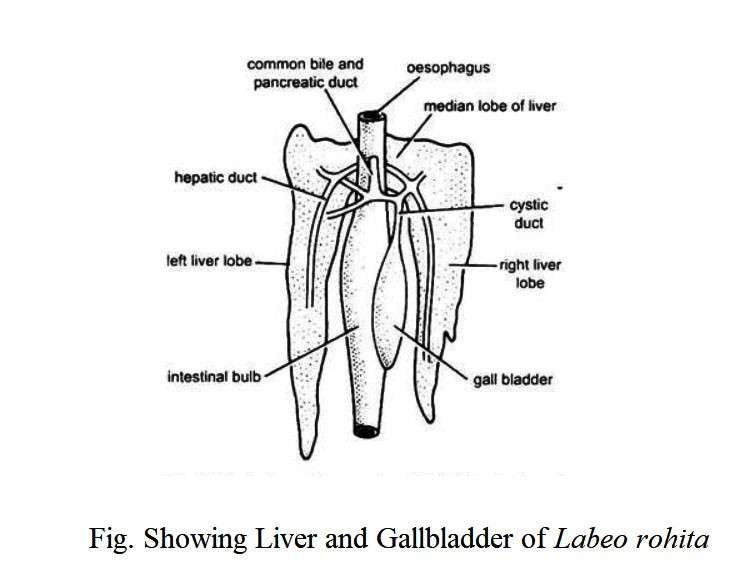
Liver: Although the liver has a variety of functions, it plays an important role as a digestive gland. The liver emerges as a hollow projection from the lower edge of the fetal duodenum. Soon the projection is divided into anterior and posterior part. The anterior part expands rapidly to form the large glandular part of the liver and the bile duct, and the cystic duct of the gallbladder is formed from the posterior part. The paired hepatic duct from each hepatic segment connects to the cystic duct of the gallbladder, forming the bile duct and is exposed to the posterior end of the duodenum.
The liver of Elasmobranch is large in size and consists of left and right segments. Both pieces are connected at the anterior end. With the exception of a few species of sharks, most elasmobracnch have a gallbladder.
Large-sized livers exist in the teleost. It consists of 2-3 segments and is attached to the anterior end. They also have a large gallbladder. In the case of dipnoin, especially in the Protopterus, there is a segmented liver located on the right ventral side of the alimentary canal. There is also a large gallbladder on the left side of the liver.
Pancreas: The pancreas is usually a dull colored soft gland that occupies the body cavity within the intestinal coil. It is the second largest gland in the body of a fish. It extends to the liver and forms the hepato-pancreas. Both histological and functional point of veiw, the pancreas is made up of two different components, one of which acts as an exocrine part and the other as an endocrine part. Their exocrine cells are large and columnar in size and they carry large nuclei. These are situated around the blood vessels or form ascini.
Each exocrine cell consists entirely of a homologous base of cytoplasm and a number of zymogen-granule rich apex. Labeo rohita, Cirrhina cirrhosus, Catla catla contains these exocrine cells in the tissue of spleen. The exocrine part secretes pancreatic juice (enzymes) for digestion and the endocrine part secretes hormones (insulin, glucagon). The primary or primitive pancreas is formed in the form of one or more buds from the ventral end of the liver and similar buds are formed from the dorsal side of the anterior intestine.
The pancreas in the elasmobranch is fully develpped from the dorsal bud of the anterior intestine. It consists of a dorsal and a ventral segment which is in most cases connected by a narrow connector. The pancreatic duct enters the intestine and releases its secretions. In adult teleost, the pancreas exists as a higher scattered gland. It is formed when the dorsal bud develops from the intestine. Some adult teleosts have a hepatopancreas and a pancreatic bud in the pancreas that forms at the junction of the liver buds. The pancreatic tissue spreads and expands in the mesentery near the anterior intestine of the adult fish. In some other fish, the endocrine material differs from the exocrine material. In the case of dipnoin, the pancreas has a distinct pervasive structure that exists in the intestinal wall.
Glands in Digestive Tracts
The mucous membrane of the alimentary canal and the sub mucosa contain the digestive glands. With the exception of some species of catfish, such glands are not seen in the mouth cavity. These fish carry fertilized eggs in their mouths. During the breeding season, the oral epithelium of these fish is temporarily folded and converted into oral glands. The secretion of these glands provides nutrition to the eggs until the larvae hatch. Such glands are missing in the pharynx. However, in a small number of teleosts (Drosoma petenense, a clupied), three is a pair of tubular projections that hang on the roof of the pharyngeal cavity. This gland is called the suprabranchial gland which helps prevent plankton from entering the water. Goblet glands exist in the glandular structure of the gastric mucosa in the cardiac and pyloric regions of the stomach. It has a simple, branched tubular structure with juice producing glandular cells.
The secretory cells of the intestinal mucosa are composed of goblets and glandular cells. The edges of the glandular cells are linear and have granular rings. There are also columnar linear cells and some plane-type columnar cells that are involved in secration and absorption, respectively. In most cases, there are no digestive glands in the rectum of the fish. The rectal gland of the elasmobranch is not the digestive gland. It releases large amounts of Nacl from the blood of marine fish.
Taste Buds and Mucous Secreting Cells: A significant number of species such as Catla catla, Puntius sophore, Tor tor, Cirrhina cirrhosus, Schizothorax plagiostomus, etc. have numerous taste buds in their beaks and pharymx. However, some fish such as Channa gachua, C. striatus, Mystus vitatus have a very small number of taste buds but other fish do not.
The taste bud may or may not be present in fish based on the fish’s diet. Many carnivorous and predatory fish (Harpodon nehereus and Muraenesox talabon) take food with the help of eyes. Taste buds are rarely seen in the mouth. Some species such as Tor tor, Catla catla, Cirrhina cirrhosus, etc. rely more or less on the taste buds for eating food. In winter, this organ contains innumerable taste buds.
The mouth cavity of all teleosts contains mucus-secreting cells that secrete large amounts of mucus and mix with food for easy swallowing. A significant number of fish have taste bud and mucus-secreting cells on the lips, oral valves and barbels for food selection.
Some herbivores and omnivorous fish such as Cyprinus carpio, Labeo rohita, Catla catla, Puntius spophore, Tor tor have tube-like esophagus. Some carnivorous and predatory fish such as Wallago attu, Channa striatus, Harpodon nehereus, Muraenesox telabon have long and swollen esophagus.
Histological point of view, the anterior region of the esophageal mucosa consists of stratified epithelium and the posterior region of columnar epithelium. Some species such as Labeo rohita, Scizothorax plagiostomus, Tor tor, Catla catla, Mystus gulio have mucus secreting cells in the mucosa and taste buds. However, it is not found in Trichiurus, Caranx and Rita rita. They have a thick layer of circular muscle fibers at the muscularis level, and longitudinal muscles spread in separate bundles on the inside.
Table: Differences between the Digestive System of Herbivorous and Carnivorous fish
|
Herbivorous Fish |
Carnivorous fish |
|---|---|
|
1. The jaws and palates of herbivores are completely toothless. |
1. These fish have strong teeth in different parts of the mouth and pharynx. |
|
2. In this case, the gill rakar is longer and thinner and the gill rakar forms a sieve-like structure along the gill openings. |
2. In this case, the gills are usually long, stiff and have tooth-like rasping limbs. |
|
3. These fish have innumerable taste buds on their beaks and buccopharynx. |
3. These fish have less number of taste buds. |
|
4. The stomachs of these fish are small in size. However, it becames too thick and forms like a gizard. |
4. The stomachs of these fish are usually pouch-shaped with thick-wall. |
|
5. Their intestines are usually long, thin-walled and more coiled. |
5. Their intestines are short and fairly straight. |
|
6. They have high levels of carbohydrate enzymes in their gut. |
6. They have low levels of carbohydrate enzymes in their gut. |
|
7. These fish have low levels of protease enzymes in their intestines. |
7. These fish have high levels of protease enzymes in their intestines. |
|
8. Example: Labeo rohita, L. calbasu, L. gonius, Hypophthalmichthyes molitrix |
8. Example: Wallago attu, Mystus seenghala, M. aor, Channa marulius, Chitala chitala, Herpodon nehereus. |
Table: Differences between the Digestive System of Cartilaginous fish(Chondrichthyes) and Bony Fish (Osteichthyes)
|
Cartilaginous fish (Chondrichthyes) |
Bony fish (Osteichthyes) |
|---|---|
|
1. The mouth is located on the ventral side. |
1. The mouths of most fish are located on the margins of the body. |
|
2. The teeth of the cartilaginous fish are scattered all over the roof of the mouth or attached to the cartilaginous jaw by the connective tissue (acrodont). |
2. The jaw teeth are located above the premaxillary and maxillary bones and below the dentine bones. Oral teeth are located on the vomar, palatine, ectopterygoid bones and tongue. |
|
3. Their teeth can be replaced. |
3. Their teeth are not replaced. |
|
4. Their teeth are similar in shape. |
4. Their teeth are usually of different shapes. |
|
5. They do not have pharyngeal teeth. |
5. Some fish have pharyngeal teeth. |
|
6. Their stomach (Elasmobranch) is “J” shaped (except: Holocephali, does not have stomach). |
6. Their stomachs are usually ‘V’ shaped (but some fish such as Cyprinus, Labeo etc. do not have stomachs). |
|
7. Scrolls or spiral valves are present in intestine. |
7. The intestine does not have a scroll or spiral valve (except Protopterus). |
|
8. The small intestine of Elasmobranch and Chimaeras is shorter than the stomach. |
8. Their small intestine is usually larger than the stomach. |
|
9. They have rectal glands in their digestive system. |
9. They do not have rectal glands in their digestive system. |
You might also read: Digestion of food in Fishes

