Labeo rohita is the important food fish that belongs to the carp family Cyprinidae under order Cypriniformes of Class Actinopterygii. It is also known as rui, rohu, or roho labeo. It can tolerate a wider temperature range, and it is the most adaptable and important cultured fish species in Bangladesh, India, Pakistan, and Myanmar.
They are column feeders and mainly feed on plant-based food. Due to their slightly downward mouth with thick lips, they feed on aquatic plants, weeds, and occasionally take the rotten organic matter from the bottom region. When they are immature, they eat plankton. They also take fish meal, mustard oil cake, rice corn, etc. as supplementary food during cultivation in ponds.
Like other vertebrates, digestive system of Labeo rohita consists of the digestive tract, and the associated digestive glands. Their digestive tract is long and twisted. The digestive tract or alimentary canal of Labeo rohita is divided into the following parts:
- Mouth
- Buccal cavity
- Pharynx
- Esophagus
- Intestinal bulb
- Intestine and
- Rectum with its external opening, the anus
Digestive Tract
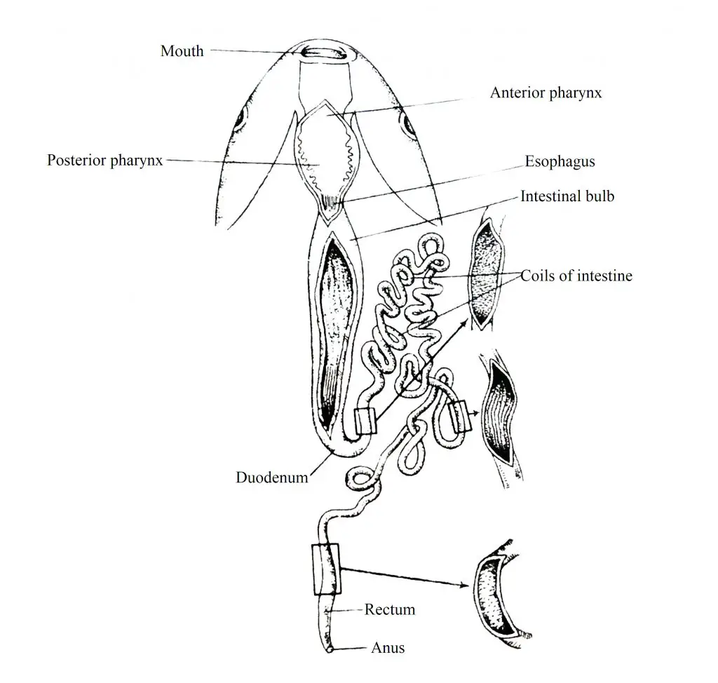
Mouth: The mouth is round and it is placed slightly ventrally at the anterior end of the head and is bounded by two soft fleshy upper and lower lips. At the free end of the lip, 4-5 rows of light black conical papillae are attached. There are no papillae in the inner folds of the lips.
Fig. Showing Different Parts of Digestive Tract of Labeo rohita
Buccal Cavity: It is a small cavity after the mouth. It is short and dorso-ventrally compressed cavity and its floor is flat and the roof is arched. On the mucous membrane of the inner wall of the buccal cavity, tiny papillae are scattered. Note that the Labeo rohita does not have a distinct tongue but the mucous membrane lining the floor of the buccal cavity which is provided with highly developed thick muscles.
Pharynx: The buccal cavity is exposed to the dorso-ventrally compressed pharynx. The pahrynx is surrounded by gill arches and it is well-demarcated into two parts. The anterior part of the pharynx is called the respiratory part and the posterior part is called the musticatory part. The lateral wall of the respiratory part is decorated with gill slits. The lateral wall of the posterior mustigatory part has 3 rows of pharyngeal teeth. When the teeth decay, new teeth are formed. They are called homodont because the teeth are identical. The base of each tooth is called the crown. The base of the tooth is embedded in the mucous membrane of the pharynx. These teeth break down complicated and solid food into small pieces. These teeth also take part in crushing food. Gill rakar is located on the inside of Gill Arch.
Esophagus: The posterior region of the pharynx leads to a very short tube, the esophagus. The wall of the esophagus is thick and consists of several longitudinal folds. The enlarged portion of the air sac (swimbladder), called ductus pneumaticus, is exposed into the esophagus.
Stomach: Labeo rohita lacks true stomach. Different scientists have different ideas about the exact cause of its absence.
Intestinal bulb: The anterior part of the intestine becomes swollen into a sac just behind the esophagus. This is called intestinal bulb. Although there are no digestive cells in the inner wall of the intestinal bulb, mucus cells and absorptive cells are present in the mucous wall.
Intestine: The intestine of a mature Labeo rohita is up to 7.5 m long. The intestinal wall is also thin and its diameter is almost the same everywhere. The whole intestine forms a number of coils. The intestinal wall also contains absorptive cells and mucus cells.
Rectum: The terminal part of the intestine swells slightly to form a thin-walled sac, called rectum. The rectum is exposed to the exterior through the anal opening, the anus. The anal opening is located just in front of the urinogenital opening. The pyloric ceaca are lacking in Labeo rohita.
Digestive Glands
The digestive glands consist of liver and pancreas.
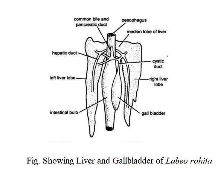
Liver: The liver of Labeo rohita (Rohu) is well-formed and dark brown in color. The gland is long and divided into two lobes. The right lobe is narrow and the left lobe is relatively wide. The two lobes are connected horizontally in two or three places. A gallbladder is located between the right and left lobes of the liver. The gallbladder is attached to the left wall of the intestinal bulb. The gallbladder is about 6 cm long and 2.5 cm in diameter. A bile duct exits from the anterior end of the gallbladder and enters the intestine. Before entering the intestine, 3 bile ducts formed from different parts of the liver join the bile duct. Bile is secreted from the liver cells and stored in the gallbladder.
Pancreas: The digestive gland, pancreas is also confined to the coil of the intestine. The pancreas extends to the liver. The exocrine part of the pancreas forms the acinus. Acinus cells are columnar and have clear nuclei. The cells are divided into basal and apical parts. Although the cytoplasm of the basal part is homogeneous in nature, the apical part carries large zymogen granules. The gland opens into the intestine through the pancreatic duct and secretes digestive enzymes.
Mechanism of Digestion
Labeo rohita are vegetarian or herbivorous species and feed on small aquatic plants, including plankton. Ingested food items are cut and lubricated with the help of existing teeth in the pharynx and temporarily deposited in the intestinal bulb. The digestion of food is done in the intestines. Digestive enzymes are secreted from the intestinal mucosa and pancreas.
The liver secretes bile into the digestive tract. The enzymes such as tipsin, erepsin, enterokinase, amylase, lipase, and maltase complete the digestion of proteins, carbohydrates, and lipids, respectively. The essence of digested food is also absorbed in the intestines. The indigestible part is stored in the form of feces and is excreted out of the body through the anus. As this fish is herbivorous, the concentration of carbohydrate-splitting enzymes is highest and protein-splitting enzymes are lowest in concentration.


