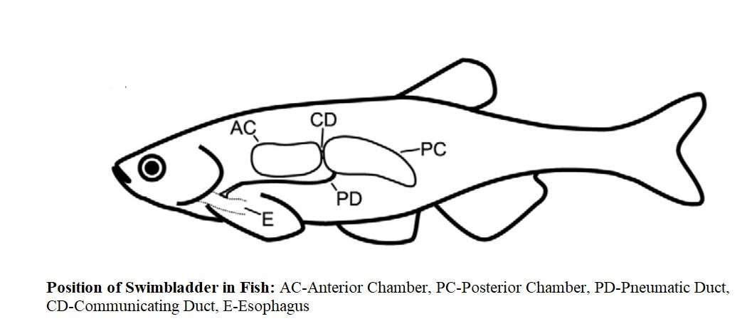
The sac-like structure between the intestines and the kidneys of most fish is called a swimbladder. However, many times it is also called gas or air sac. It contains a mixture of carbon dioxide (CO2), oxygen (O2) and nitrogen (N2) gases. With the exception of a few areas, almost all bony fish contains swimbladder. The swimbladder usually opens in the esophagus through a duct called the ductus pneumaticus. This duct is shorter and wider in the lower bony fishes (Chondrostei and Holostei), but in other cases, it is longer and narrower.
Pneumatic duct is a special type open duct in the case of some classes of teleosts such as Clupeiformes, Esociformes, Anguiliformes and Cypriniformes. Dactus pneumaticus is absent in Gasterosteiformes, Mugilliformes, Notacanthiformes and Acanthopterygii. Therefore, bony fish are divided into physostomi with open ductal swimbladder and physoclisti with closed swimbladder.
In some cases, the absorption and secretion of swimbladder cannot be subtly separated from each other. Such swimbladder are called paraphysoclists. On the other hand, some fish have different absorption and secretory zones. Such swimbladder are called euphysoclists. In higher vertebrates, the position of the swimbladder is similar to that of the lung. It is considered to be homologous part of the lungs. However, the difference between the two is mainly in the process of origin and blood supply.
The swimbladder originates from the dorsal wall of the alimentary canal and receives blood supply from the dorsal aorta. The lungs, on the other hand, originate from the ventral wall of the pharynx and receive blood supply from the 6th carotid arch. In most bony fish, swimbladder act as hydrostatic organ.
Development of Swimbladder
There is disagreement about the development of the swimbladder. In the case of teleost, it originates as a diverticulum resembling the dorsal or dorso-lateral sac of the esophagus. The sac is connected to the esophagus by an openimg. Later it was divided into two halves. Except for some fish, the left half is eroded. The right half is well developed and takes the middle position.
In the case of Dipnoi and Polypteridae, it is formed from growth in the lower part of the pharynx and transforms into a lung. This growth turns to the right and is located on the dorsal side of the alimentary canal. As a result of such a transfer, the right lung moves to the left. According to Spengel, the swimbladder originates from the posterior end of the gill pouch. However, embryological support was not found.
Fundamental Structure of Swimbladder
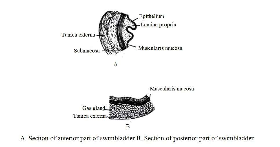
In the case of different fish, the structure, size and shape of the swimbladder varies. It is a strong sac-like structure with a capillary blood vessels on the wall. Beneath this capillary blood vessels, a layer of connective tissue called tunica externa. Below this layer, there is a layer of smooth muscle and epithelial gas glands called tunica interna.
The swimbladder has one or two or more chambers. In the case of teleost, it is located between the intestine and the gonad below the kidney. There may be a lifelong connection with the esophagus or it may become extinct in the mature state. According to Biot (1806) and Moren (18), most of the gas emitted by swimbladder is oxygen. The rest contains nitrogen and a small amount of carbon dioxide.
Types of Swimbladder
Swimbladder can be divided into two types based on the presence of ductus pneumaticus between the swimbladder and esophagus, namely: Physostomous and Physoclistous. Moreover, in eel fish (anguilliformes), transition condition of swimbladder between these two types is seen.
Physostomous
When there is ductus pneumaticus between the swimbladder and the esophagus, it is called physostomous. A blood vessel arising from the ciliaco-mesenteric artery supplies blood to the swimbladder and the blood returns to the heart through the hepatic portal vein. Such swimbladder can be seen on bony ganoid fish, lung fish and soft ray-finned teleost.
Physoclistous
In this case, ductas pneumaticus is closed or like a bud. A type of swimbladder can be seen in spinus rays finned fish. In this case, the swimbladder consists of the secretory gas gland (rete merabilia) on the anterior side and the ovale gas absorbing region on the posterior surface. The oval develops from outside of the eroded ductus pneumaticus.
The ciliaco-mesenteric artery and the dorsal aorta supply blood to the gas gland of Rete mirabilis, the oval, and the wall of the swimbladder. On the other hand, blood from the gas glands returns to the heart through the hepatic portal vein and blood from the remnants of the sac returns to the heart through the posterior cardinal vein. The sac, especially the gas gland, is nerved from a lateral branch of the vagus nerve and by the oval sympathetic nerve.
Transition Phase
The transition condition of physoclistous and physostomous is seen in eel fish (Anguilla). In this case, the ductus pneumaticus of the swimbladder expands and forms a distinct oval chamber. There is also a gas gland in such a sac that receives blood from a branch of the ciliaco-mesenteric artery to the swimbladder and carries blood to the heart through the post-cardinal vein. In the evolution of the swimbladder, this condition occurs when the physostomous are transformed into physoclistous.
Swimbladder and Its Modifications
Wide variations are observed in the size and function of the swimbladder of fish. Elasmobranch, bottom-dwelling and deep sea teleosts have an intermediate bud-like swimbladder during development period but do not remain in a mature state. Flat fish (Pleuronectidae) have swimbladder in the early stages of life when they are stayed in the pelagic position, but when they reach adulthood and live lazily at the bottom, the swimbladder is eroded.
According to Miklucho-Maclay (18), in the embryonic stage of Squalus, Galeus and Mustelus, the dorsal diverticulum of the bud is seen from the anterior alimentary canal. Most teleosts have swimbladder that has been adapted to a variety of life styles.
Modifications of Physostomous Condition
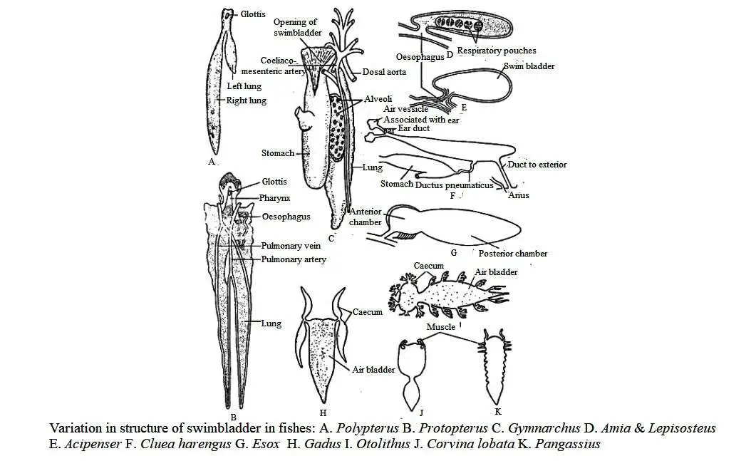
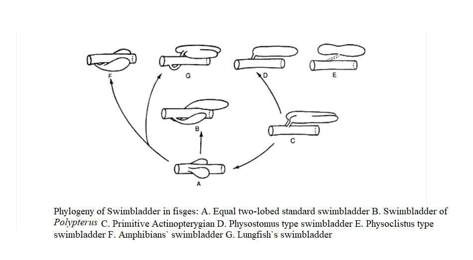
In some species, the ideal phasostomous are transformed into twin sacs. As a result, it becomes the front and back chambers. In the case of chondrostei, especially in Polypterus, the swimbladder is of the primitive type, it is divided into two unequal chambers, the left lobe is short and the right lobe is long and the sac is connected to the pharynx by the glottis.
The glottis has muscular sphincters. The inner surface of the sac is smooth and partially ciliary and does not contain a co-alveolar sac but has a muscular wall. A pair of pulmonary arteries originating from the last pair of epibranchial arteries supply blood to the sac and the corresponding vein enters the hepatic vein and the blood returns to the heart through the sinus venous. In the case of Acipenser, it is small and oval.
The ductus pneumaticus enters the ventral side of the sac and is exposed to the intestine on the posterior side of the pharynx. In this case, there is no glottis. The opening of the pharynx are closed by a contraction of the ductus pneumaticus. The outer wall of the sac is fibrous, thick but the inner wall is smooth. In this case, the right and left lobes are formed from the dorsal side of the esophagus in the embryonic stage. However, the left lobe becomes extinct and from the right lobe, a mature swimbladder is formed.
In the case of lungfish (Dipnoi), the inner wall of the pouch becomes rich in numerous alveoli and turns into lungs. This sac is structurally and functionally similar to the lungs of tetrapods. In Neoceratodus, the sac has one lobe but in Lepidosiren and Protopterus it has two lobes.
In the case of Holostei, especially in Amia and Lepisosteus, a swimbladder is formed in which an inconspicuous sac expands along the entire body cavity. In both cases, the left lobe looks like a bud in the embryonic stage but it exists for a short time. The ductus pneumaticus is exposed to the esophagus by a dorsal opening-like glottis at the back of the pharynx.
The sac wall is rich in capilary blood vessels and contains pulmonary alveoli. Swimbladder of Amia receives blood supply from the pulmonary artery and returns to the heart from the left Ductus cuvieri, whereas in Lepisosteus it receives blood from the dorsal aorta and returns blood from the right post-cardinal vein.
In the case of teleostei, especially in Gymnarchus, a transition condition of the Amia and Lepisosteus is seen. In this case, the efferent branchial artery from the third and fourth gill arch connects to form a common blood vessel from which the pulmonary and ciliacomecentric arteries originate. The ductus pneumaticus of Clupea harengus is attached to the fundus region of the stomach and another duct in the posterior part of the swimbladder is exposed to the anus.
Similar posterior opening exist in Pellona, Caranx, Sardinella. The size and spape of the swimbladder varies greatly. In some cases, it produces numerous branched projections (diverticula). In many fish, the anterior growth of swimbladder comes in close contact with the inner ear wall. In the Clupea, the narrow front of the swimbladder enters the baso-occipital duct of the skull and splits into two narrow branches, the front end of each branch swelling to form a round bulge that comes in contact with the inner ear.
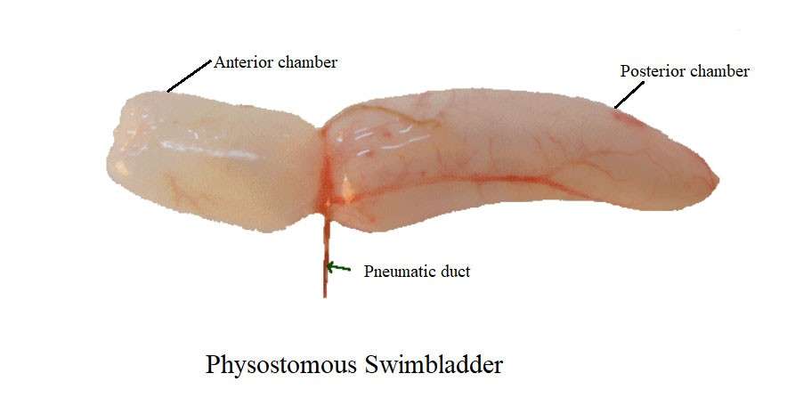
More or less similar conditions are seen in Tenualosa ilsha. Finger-like projections from swimbladder are seen to be formed in many fish. In Gadus, a pair of projections is generated from the tip of the swimbladder and enters the head area. The swimbladder is transversely divided into anterior and posterior chambers in most species such as Cyprinoids, Esox, Catostomus, Pungassius etc.
In the case of Arius, it splits longitudinally. In the case of Notopterus, a longitudinal membrane divides the notch into two lateral chambers. Due to the presence or absence of a membrane, the inner chamber of the swimbladder is completely or partially divided.
Modifications of Physoclistous Condition
In all teleostei, the swimbladder begins in the physostomous state but in the mature stage the ductus pneumaticus is eroded and turns into the physoclistous stage. An ideal physoclistous swimbladder is a closed sac with anterior and posterior chambers. Chambers are connected by an opening called two ductus communicus. The opening and closing of these pores are controlled by sphincter-like circular and radial muscles.
The bud stage of the swimbladder enlarges to form the anterior chamber and the ductus pneumoticus expands to form the posterior chamber. The ideal physoclistous phase has been modified differently in different species. The rete merabilia of the posterior chamber becomes flattened and it is known as the oval. This condition is seen in Myctophidae, Percidae, Mugilidae etc. The oval reabsorbs gas and its openings are regulated by circular and longitudinal muscles. Such techniques play an important role in the rapid vertical movement of fish.
Histological Modifications of Swimbladder
In different species of fish, anatomical modification of swimbladder occurs due to modification of histological modification. The fish’s swimbladder acts as a hydrostatic organ. It helps the fish to sink or go to different depths of water by changing the gas holding capacity of the sac. Fish with open ductus pneumaticus can change gas holding capacity by inhaling and exhaling air. On the other hand, in some physostomous and all types of physoclistous species, this gas is transferred from the bloodstream. In this case, there is an oxygen producing and absorbing system in the sac.
Swimbladder is rich in blood vessels, but the amount of blood vessel varies from species to species. In some species of the family Clupeidae and Salmonidae, the blood vessels of the swimbladder is equally rich throughout, but in most cases, the blood vessel is densely embedded to form rete mirabilis. On the other hand, other physostomes such as carp (Cyprinus, Labeo, Tor tor), blood vessels are arranged like fins and condense at one or more points of the swimbladder to form red lumps of different shapes. This gland is called red gland. This gland is located in the anterior chamber of the swimbladder, which contains oxygen-secreting cells.
Blood from the ciliaco-mesenteric artery enters the red gland and travels back through the portal vein. The function of this gland is controlled by the vagus nerve. Species that do not have functional ductus pneumaticus, they do not have gas glands, but in the case of eel fish, the red glands do these functions. In Physoclistous species, the anterior region of the swimbladder is adapted to gas production and the posterior region involves to gas absorption.
As the wall of the back chamber is too thin, diffusion of gas occurs easily. Gas is absorbed by the blood under the wall of the swimbladder. Some fish have a gas-absorbing region called the oval located on the back surface of the swimbladder. Oxygen can easily enter the capilary blood vessel through the epithelial lining of the oval. The gas-absorbing region receives blood from the dorsal aorta and the blood returns through the postcardinal vein. This process is controlled by the sympathetic nerve.
Histological differentiation for gas production and absorption is an important achievement in the case of fish. The gas that is produced from the red glands is mainly oxygen. This oxygen is rapidly absorbed or permeated in the capilary blood vessels. In the alternative process, gas production and absorption is done by increasing or decreasing the internal pressure and gas holding capacity of the sac. The red gland is usually located in the anterior chamber, but in the case where the anterior chamber of the swimbladder is involved in hearing function, the gas gland is located in the posterior chamber.
From a histological point of view, the wall of anterior chamber of the swimbladder of fish under the family Cyprinidae consists of the following parts:
(1) Tunica external – It is composed of concentrated collagen fibrous material.
(2) Submucosa – It is made up of loose connective tissue.
(3) Muscularis mucosa – It consists of a thick layer of smooth muscle.
(4) Lamina propria – It consists of a thin layer of connective tissue.
(5) Inner most layer – It is made up of epithelial cells.
Different parts of the posterior chamber of the swimbladder may have internal epithelium with different heights and shape. The outer layer of smooth muscle contains a glandular layer of fine-grained cytoplasm with large and irregular cells (Fange 1955). This glandular layer receives adequate blood supply from the rete mirabile. Such blood vessel also exists outside the muscle layer.
The ductus pneumoticus connects the two chambers of the swimbladder and contains nerve fibers at its strong muscle level. The idea is that the muscles of the posterior chamber of the swimbladder have a controlling effect on the gas glands and it also controls the volume of the swimbladder. The muscles of the ductus pneumaticus act as a sphincter.
Relation Between Swimbladder and Auditory Apparatus
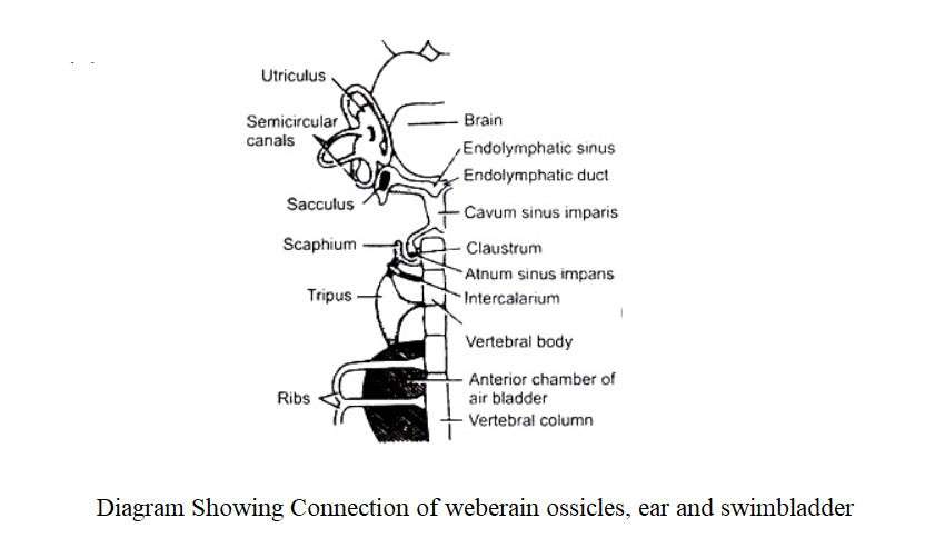
In some fish, the swimbladder is closely related to the inner ear so that changes in perilymph pressure can be easily transmitted. Evidence of its evolution is found in the connection of the swimbladder and the auditory instrument. Gadus sp, Megalops sp and other fish have this simple type of connection.
The finger-like diverticulum arising from the tip of the swimbladder connects to the auditory capsule through an opening by the membrane covering. In fish of the family Clupeidae, the connection is more intense, in which case, a pair of tubular growths enter the auditory capsule. Each branch divides again and ends in a single swelling vesicle.
These vesicles cover the pro-otic, ptero-otic bones and connect to the membrane labyrinth. In the case of carp (Cyprinidae) and siluriformes, this connection is further enhanced by the fact that it is connected through a bone chain called the Weberian ossicle. This Weberian ossicle is formed by the transformation of several anterior vertebral segments.
Functions of Swimbladder
Swimbladder of fish does different functions. Its functions are mentioned below:
1. As Hydrostatic Organ:
It first acts as a hydrostatic organ. It equals the weight of the body with the weight of the water removed. The density of fish meat is 1.076 and the density of fresh water is 1.0005 and the density of salt water is 1.026. Swimbladder can increase or decrease the overall weight of the fish by generating or absorbing gas. As a result, fish can float anywhere in the water. They have oil and fat in their muscles to lighten the body weight of the fish. Fish swimbladder accounts for 4-11% of total fish volume. In case of marine fish it is 4-6% and in case of freshwater fish it is 7-11%.
2. As Respiratory Organ
Swimbladder of many physostomous fish act as temporary or complementary respiratory organs. The swimbladder of the Amazon giant red fish (Arapaima gigas) is adapted for aerial breathing. In addition, swimbladder of Bichir (Polypterus), Boffin (Amia), Lepisosteus and Notopterus help in aerial respiration. When the water temperature reaches 200 C, the bichir fish comes to the surface for air.
The swimbladder of the lungfish resembles the lung of amphibians. They breathe through their lungs during aestivation. All of these fish travel to the surface of the water to take in oxygen through mouth gap and release carbon dioxide. Since swimbladder of physostomous fish contains more carbon dioxide gas than air, as a result, waste gas carbon dioxide is emitted from here.
3. As Auditory Organ
Any change in the aquatic environment around the fish actually enters the lateral organ in the form of water waves. These are received by the neuromast cells of the lateral organ which reach the swimbladder through the lateralis branch of the vagus nerve and then enter the inner ear. In this context, the fish is able to take defensive measures.
4. As Sound Production
Records have been found of fish sound production by placing hydrophones at different depths of water. Out of 20,000 fish species, only a few hundred species are capable of producing sound. Some fish such as Drums (Sciaenidae), Granadiers (Melanoridae) and Gurnard (Triglidae) etc. are capable of producing sound. In this case, striated muscles of the dorsal body wall and the inner wall of the swimbladder help to produce sound.
In toadfish (Batrachoididae), the striated muscles of the swimbladder can produce audible signals by rapidly changing their size. The sound generated by the swimbladder is of low frequency. Fish attract their mate by making sound in reproductive behavior. Besides, it also plays a role in protecting the reproductive area. Thus the Minnow (Cyprinidae), Loaches (Cobitidae) and Eel (Anguillidae) fish are able to generate sound by rapidly expelling air from the swimbladder through pneumatic ducts.
Herring (Clupeidae) produces sound by emitting gas from the swimbladder through the duct at the back of the anus. In the case of deep-sea fish, which do not have biological luminosity, sound production plays an important role in maintaining species-to-species communication.
5. As Sound Receptor
The concentration of fish is close to brackish water. The bone and swimbladder work together as sound transmitter or resonators. Some fish have swimbladder extending to the inner ear. Differences in pressure due to sound waves are transmitted directly to the perilymph, as in the case of cod (Gadidae), Porgies (Sparidae), the outgrowth of the anterior edge of the swimbladder touches the cranial bone of the inner ear of saculus.
The sound waves reach the inner ear by changing the size of the swimbladder in the fish under order Cypriniformes. In this case, the Weberian ossilcle connects the swimbladder and the inner ear. Sound-receiving sensory cells are located below the labyrinth. In the case of fish under order Cypriniformes, the size and shape of the swimbladder varies from family to family.
The swimbladder of Minnows (Cyprinidae) is divided into anterior and posterior chambers. In the middle of both chambers, there is a sphincter of smooth muscle. In the case of bottom-dewlleing Cypriniformes fish such as Loach and others (Homalopteridae, Gastromyzonidae I Cobitidae), the posterior chamber of the swimbladder is often extinct and the anterior chamber is located in the bony capsule. There is jelly-like liquid here.
Many catfish (Sisoridae) have only one anterior chamber that moves through the capsule and comes out of the skin through the lateral opening of the capsule. They can do feel the difference in sound more than fish without swimbladder. Some Minnows (Cyprinidae) can detect differences in water pressure up to a few cm. In this way, they can also understand the fall of rain or the decrease in rainfall and the change in the level of dissolved oxygen.
Role of Swimbladder in the Distribution and Ecology of Fishes
In the case of modern fish, various mechanisms have emerged to prevent buoyancy. Although the body density of fish is equal to the density of water, extra energy must be expended to maintain their position in the flowing water. Many river darters (Etheostomatinae) do not have swimbladder or air sac. However, swimbladder exists in Log perch (Percina carprodes) of the same subgroup.
The extinction of the posterior chamber of the swimbladder has occurred in the Loach group living in the mountainous flowing River in South Asia. In all loaches, the front of the swimbladder connects the two chambers to the inner ear, which is used as a hearing aid. From this relationship, it is known that in rapid currents, the swimbladder automatically and individually leads to extinction. However, when one part is lost, the fish moves from the deepest part of the ocean to the top with the highest development of other other part.
It has shown from the Trolling that the number of Mesopelagic fish is higher between 200-1000 meters. There are so many of them here that the sound is made by Sonar in the sea. The sound-making fish are seen coming to the surface at night and the fish go deep during the day. These echoes are reflected features of small gas bubbles and are thought to reflect the sound waves of the swimbladder of fish.
Mesopelagic species, especially lantern fish (Myctophidae), migrate to the surface of the ocean by vertical migration for plankton food at night. Their predatory fish such as deep sea viper fish (Chauliodus) and deep sea squawlower (Chiasmodon) make extensive vertical migrations. Many mesopelagic species develop capillary blood vessel or oval organs to reabsorb gas-emitting complexes.
Air sacs of Mesopelagic fish are more advanced than groups that have rete mirabile or gas glands. Like the Flyingfish (Exocoetidae), on the surface, the gas-emitting complex of swimmers is about 1/10 of the total air sac volume, while the Lantern Fish (Myctophidae) and other mid-depth fish are more extensive. Due to the greater expansion of the gas exchange structure, the mid-depth fish make massive migrations (3-400 m), they rise on the surface at night and go down again in the morning.
The gas sacs of some fish of the family Melamphacidae in the lower pelagic region are filled with fat. Like lantern fish they cannot migrate as extensively. However, it lightens the muscles and bones of the fish. Roughly filled gas sac with fat do not play a significant role in lightening the fish.
More than deeper oceans of 1000-4000 meters are not conducive to living. The fish here do not have any gas sac or swimbladder. Bone loss, muscle weakness occurs due to more water here. The animals of this region are lazy in nature and move a little. The concentration of gas is high due to the excessive pressure of water on the surface of the fish
In the Bethyal, Abyssal and Hadal areas of the ocean floor, where it is 2000-5000 meters or more below, food is stored again. As a result, benthic invertebrates and fish are found. Here 50% of fish have an air sac or swimbladder. They have heavy bones and strong muscles. The blood vessels of rete mirabile are longer than the swimbladder. It is thought that these fish in the deepest areas, swimbladder helps in the search for food.
Physoclistous fish (Anguillidae and Microstomidae) predominate in the marine environment. Freshwater physostomous fish such as salmon (Salmonidae) have consistently lost the actual connection between the swimbladder and the esophagus. Many physoclistous fish are found in freshwater but physostomous fish are predominant in freshwater fish.
Mechanism of Filling and Emptying of Swimbladder
However, the partial pressure of oxygen and nitrogen in the water of most natural reservoirs and in the blood of arteries of fish is 0.2 and 0.8 atmospheric pressure, respectively. On the other hand, the oxygen and nitrogen pressures of the swimbladder are 100 and 20 atmospheric pressures respectively. The swimbladder is the only organ that is able to concentrate oxygen 500 times and nitrogen 30 times.
With the loss of the yolk sac, most physostomous fish fill the air sac or swimbladder by swallowing the air. Although these fish can expel and absorb gas through the blood supply, in adulthood they are not able to fill the swimbladder without the help of the atmosphere. Many physoclistous fish such as Stickleback (Gasterosteus), Guppy (Lebistes), Sea horse (Hippocampus) etc. have pneumatic ducts in the larval stage. At this time they fill the sac with air from the atmosphere.
Fossil records of Lobfin (Crossopterygii) fish and the presence of pneumatic ducts in the larvae of Physoclistus fish prove that the primitive bony fish were of the actual physostomous type. Any deep-sea fish has a functional gas sac or swimbladder and it is thought that they live in deep water (Melanonidae) for whole life. When lantern fish (Myctophidae) are in the early stages, they take up oxygen from the surface of the water. When their initial stage is over, they go to the bottom and adopt various techniques to fill the swimbladder or air sac.
The pressure and volume of the air sac or swimbladder increases or decreases as the fish moves up and down as needed. Due to the hydrostatic pressure, the air holding capacity of the air sac or swimbladder of fish is fairly stable. According to Boyle’s law, changes in gas pressure also result in changes in volume, which also applies to air sac. Gas emission and absorption is a biological process and there are limitations to the speed of the fish in moving with depth.
The European perch (Perca fluviatilis) can climb up to 20 meters at ease and can do feel sound within 18-20 meters. This species can move up to 20% of the total depth when moving from low to high. If the oxygen tension in the body of the fish is higher than in the body of water in which the fish live, then the epithelial cells of the gas gland carry the dissolved oxygen from the capillary blood vessel to the air sac. This causes a small intracellular gas bubble to enter the water. Although they can emit bubbles, the glandular cells are impermeable to dissolved gas. They prevent the movement of gas from the outside of the air sac into the capillary blood vessel of rete mirabile of the epithelium as a barrier to gas diffusion.
Air sac of deep sea white fish (Coregonus) contains pure nitrogen. The air sac of deep-water fish is usually filled with oxygen. Conger eel (Conger) increases oxygen by 16% at 1 m depth, 50% by 6 m depth and by 6% at 165 m depth. The reabsorption of gas from the swimbladder is done in several processes, viz
1. Diffusion occurs in blood vessels of the wall of the swimbladder and outside it contains gas-emitting complexes [Sauries (Scombreocidae) and Killifishes (Cyprinodontidae)].
2. It is sometimes associated with a gas-emitting material through a specialized absorbing capillary blood vessels. When the fish rises to the top, it expands and becomes fully functional. During this time the gas glands fold to absorb the gas quickly and then exit through the gills. When the fish goes down, the gas glands expand and the re-absorbing capillary blood vessel closes. The area adjacent to the gas gland increases (Sternoptychidae) due to the rapid release of gas inside the sac by the gas gland.
3. The capillary blood vessel complex is separated from the cavity of the air sac by a single-celled epithelium. In this region, the blood vessel comes from the intercostal artery and travels to the cardinal vein known as the oval organ. This organ is surrounded by a sphincter that controls the rate of gas absorption by contraction and expansion of the oval pores in the capillary blood vessel region.
Cods and their close relatives (Gadiformes) and many finned fish (Acanthopterygii) have oval organs. From an embryological point of view, the pneumatic duct of the physostomous and the oval organ of the physoclistous are congruent. The swimbladder is nerved by the branches of the vagus nerve and the ciliac ganglion. Nerve endings occur in secretory cells of the reabsorption region, the oval, the rete mirabile, and the epithelium.
Rete mirabile has numerous large ganglionic cells near the poles of the heart. Nerves also exist at the muscular level of the wall of the swimbladder, but the cause of their presence is not yet fully known. The reabsorption of gas is stimulated by catecholamines whereas in the absence of such stimulation the gas storage rate increases.
Lantern fish (Myctophidae) and other deep-water fish come to the surface at a distance of 400 meters or more vertically at night. This is why large air sac has the structure to emit and absorb high levels of gas. Moved from the cavity of the air sac, the blood vessels in the wall of the sac merge with the particular blood vessel to form two parts of the gas secretory complex.
Special vascular gas secretory region consists of two parts such as:
(1) Gas gland– Gas gland is a special sac which is a region of epithelium which consists of several layers of epithelium which are single-layered, folded or stratified; and
(2) Rete mirabile-Rete mirabile or capillary blood vessel and veins formed in the wall of the swimbladder. In Physoclistous fish it receives blood supply from the ciliaco-mesenteric artery and exits through the branch of renal portal vein. Eel fish (Anguillidae) have more than 100,000 arterioles and a small number of venules, increasing the total surface area of Rete mirabile by more than 2 square meters. The blood in the arteries of the swimbladder and the blood in the veins are mixed through diffusion.
Relationship Between Swimbladder and Lung
The fish`s swimbladder and lungs of tetrapods are similar in structural and developmental aspects. Both are attached to the lining of the alimentary canal and both originate from the growth of the galate and their glottis is in the same position. However, due to the scarcity of connecting fossil records, it was not possible to find the cause of such similarities. The information available on embryological and comparative morphology is even more confusing.
Fish`s swimbladder has evolved from simple to complex. In this case, it has finally acquired a lung-like structure in terms of structure and function. In the case of lung fish and bichir, the swimbladder actually acts as an aerial respiratory organ. Sturgeon and many other fish have primitive type of swimbladder. In this case, the swimbladder is a simple sac-like structure filled with a mixture of gases which acts as hydrostatic organ. However, the inner wall of the swimbladder of Amia, Lepisosteus, and Lungfish is enriched with more blood vessel and the surface volume is increased due to the development of pulmonary
In most cases, the swimbladder has a single structure, but in the case of Amia, Lepisosteus, Protopterus and Lepidosiren, the swimbladder is joint (it is unequal in Polypterus). The right side of the swimbladder is longer and the left side is structurally shorter and is attached to the galate through the glottis. In the case of lung fish, the arrangement of blood vessels in special organs is similar to that of amphibians. From the above discussion it appears that the origin, structure and function of the swimbladder and lung of tetrapods are similar. Due to this close resemblance, many researchers believe that the swimbladder is the forerunner of the tetrapod`s lungs in the phylogenic history of vertebrates. However, no geological support was found for this idea.
The presence of the lungs from a geological point of view is more ancient than that of a fish`s swimbladder. As a result, the idea of the origin of the lungs directly from the swimbladder is discarded. However, the idea of a single phylogenic origin in this regard has become more popular. The idea is that placoderms are more primitive nathostomes that carry swimbladder.
Both the lungs of tetrapods and the modern fish swimbladder have evolved to be the source of the placoderm. This is easy to explain, but the actual facts about evolution are insufficient. There are different opinions in this regard. Recently, it has been speculated that the swimbladdder originated from a pair of pouches that later evolved into a pouch. This condition is seen in fish in which the swimbladder acts primarily as hydrostatic organ.
The swimbladder of Polypterus, Protopterus and Lepidosiren is primitive in nature due to its transformation into lungs. Therefore, the respiratory swimbladder of the lung fish cannot be the ancestor of the teleost’s hydrostatic type of swimbladder. In this case, it is thought that the hydrostatic type of swimbladder in fish evolved individually. Amphibians have originated from the Crossopterigian ancestor. As a result, in the light of current knowledge, it is difficult to determine the actual relationship between the swimbladder of fish and lungs of tetrapods.
Difference between Swimbladder and Lung
Although both the swimbladder and the lungs play a role in respiratory function, some differences are observed between them. Only in Dipnoin fishes, lungs exist instead of swimbladder. Moreover, tetrapods also have lungs. The difference between the lungs and the swimbladder is mentioned below:
|
Swimbladder |
Lung |
|---|---|
|
The swimbladder originates from the ventral side of the alimentary canal (Polypterus) or from the lateral aspect (Dipnoi) or from the dorsal-ventral side (Teleosts). |
It originates from the ventral side of the pharyngeal wall. |
|
It is usually used as hydrostatic organ. Rarely used for respiration. |
The main function of the lungs is respiration. |
|
It is found in fish (except: Lung fish). |
It is found in terrestrial vertebrates or tetrapods. |
|
It contains O2, CO2 and H2 gases when dissolved in water. |
Only O2 enters the lungs and CO2 is expelled. |
|
It receives blood supply from the dorsal aorta. |
It receives blood supply from the 6th aortic arch. |
|
The swimbladder is divided into physostomus, physoclistus and transition. |
There is only one type of lung. |
|
The swimbladder is said to be the ancestor of the lung. Its structure is relatively simple. |
The structure of the lungs is more advanced. |
Difference Between Physostomous and Physoclistous Swimbladder
Physostomous |
Physoclistous |
|---|---|
|
Such swimbladder are connected to the esophagus by ductus pneumaticus. |
Such swimbladder does not have ductus pneumaticus. |
|
In this case, there are no gas absorbing regions such as gas glands and ovale. |
In this case, there are gas absorbing regions such as gas glands and ovale. |
|
In this case, blood is supplied to the swimbladder through an artery coming from the celiaco-mesenteric artery, and the blood returns to the heart through a vein connected to the hepatic portal vein. |
In this case, the blood supply is obtained to the rete mirabile of gas glands, oval and wall of swimbladder through the celiaco-mesenteric artery and dorsal aorta. Blood from the gas glands travels back to the heart |
|
This type of swimbladder is available on ganoid fish, lung fish and soft-ray finned teleost |
Such swimbladder is found in fish with spiny ray finned fish. |
|
In this case, the gas enters and exits the swimbladder through ductus pneumaticus. | In this case, the gas enters and exits the swimbladder through diffusion. |

