The body needs some essential foods for maintaining proper growth. These foods should be some protein like meat, fish, eggs, milk, pulse, etc. which are hydrolyzed into amino acids during digestion. Some amounts of amino acid are utilized in metabolic process of energy production. During the metabolic process, some nitrogenous wastes are produced as byproducts which are harmful to health. In this case, human excretory system (kidney) plays an important role to discharge the waste products from the body.
Urinary system is the main excretory system in humans. It performs the functions of excretion, the bodily process of discharging wastes. Besides it, the skin can also perform excretion by discharging water, small amount of urea and salts. The excretory system of human includes the following four structures:
- Kidneys
- Ureters
- Urinary bladders and
- Urethra
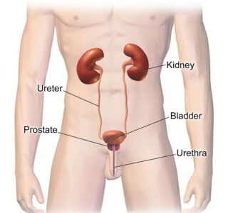
Image Showing Parts of Human Excretory System: Image credit: wikipedia.org
You might also read: Epithelium: Definition, Characteristics, types and Functions
Structure of Kidney
The human body and other vertebrates possess a pair of kidneys which are present at the dorsal side of the abdomen on either side of the vertebrae of the lumber region. The upper parts of the kidneys are partially protected by the eleventh and twelve ribs. Each kidney is bean-shaped and red brown in color.
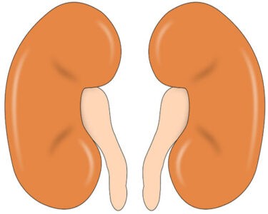
Image Showing Human Kidney: Image Credit-needpix.com
Table showing the weight of kidney:
|
Kidney type |
Male |
Female |
|---|---|---|
|
Right kidney |
80-160 gm |
40-175 gm |
|
Left Kidney |
80-175 gm |
35-190 gm |
The weight of the each adult`s kidney ranges between 80 and 175 gm in male and between 40 and 190 gm in female. The right kidney is slightly smaller than the left kidney. The kidney of an adult man is approximately 11-12 cm in length, 5-6 cm in wide and 3 cm thick. Each day, the kidneys can filter about 180 liters of blood.
The kidneys remove toxins, medications, urea, excess ions and form urine. It plays an important role to make balance salts and water as well as well acids and bases.
Internal Structure of Kidney
Each kidney has an indentation on the inner side called the hilum. Nerves, ureter and blood vessels enter the kidney at the hilum region. If a kidney is cut through the middle in two longitudinal halves, two regions are revealed to naked eye-an outer cortex and inner medulla each of which lodges different components of the kidney. It is seen that the cortical tissue penetrates the whole depth of the medulla thereby dividing it into a number of conical structures called pyramids.
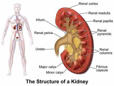
Image Showing Internal Structure of Kidney: Image credit-wikimedia commons
Internally each kidney has two distinct regions, an outer renal cortex and inner renal medulla. The renal cortex is reddish color and has a smooth texture and it is the location of the Bowman`s capsules, the glomeruli, the proximal and distal convoluted tubules and their associated blood supplies.
The renal medulla is the central part of the kidney. This area is a striated red-brown color. There are approximately 8-18 striated triangular structures called renal pyramid within the renal medulla of each kidney.
The broad base of each pyramid remains in contact with the cortex and the round apex project to the renal pelvis. The apex of two or three pyramids combines and form renal papilla. It is gradually become a tubular structure called calyx minor. Several calyx minors united to form a greater calyx major which opens into a funnel shaped cavity, the renal pelvis. Pelvis is the upper expanded portion of the ureter. The renal pelvis receives the urine drained from the kidney nephrons via the collecting ducts and then the papillary ducts. The extended parts of cortex in between the renal pyramids are known as the renal column.
Each kidney is provided with a million tubular excretory units called nephrons. Whole kidney is surrounded by a peritoneal membrane called tunica fibrosa or renal capsule made of connective tissue.
You might also read: Best Anatomical Skeletons
Ultra-structure of Kidney
Nephrons are the functional and structural units of the kidneys. Each kidney in the body contains about 900,000 to 1.2 million nephrons. A single nephron is about 5 cm long and made up of the following two distinct parts:
- Bowman`s Capsule and
- Renal Tubules
Types of Nephron: According to the position of Bowman`s capsule with the cortex, the nephrons are of the following three types:
1. Superficial cortical nephrons– In these type of nephrons, the Bowman`s capsule occupy the superficial part (outer most part) of the cortex. These comprise about 85% of the nephrons in the kidney. They have short loop of Henle.
2. Mid Cortical nephrons– Renal corpuscles are located in the midcortex, deep to superficial cortical nephrons but above the next category. These nephrons may be either short-looped or long-looped. These comprise about 5% of the nephrons in the kidney.
3. Mid Cortical nephrons– Renal corpuscles are located in the midcortex, deep to superficial cortical nephrons but above the next category. These nephrons may be either short-looped or long-looped. These comprise about 5% of the nephrons in the kidney.
Histological Structure of Nephron
There are the following two parts of a nephron:
- The renal corpuscle or the Malpighian body and
- The renal tubule
1. The renal corpuscle or the Malpighian body: The proximal globular structure of nephron is situated in the cortex region of the kidney, known as the renal corpuscle or the Malpighian body. Each renal corpuscle has the diameter of 0.2 mm and has two parts:
- The Glomerulus and
- The Bowman`s capsule
Glomerulus: The renal artery gives rise to a number of afferent arterioles. Each afferent arteriole after entering into the Bowman`s capsule divides into about fifty capillary loops. These loops form a tuft, known as glomerular tuft. It is situated in a cup-shaped structure of nephron, termed as Bowman`s capsule. The capillary loops, afterwards reunite and form the efferent arteriole. This efferent arteriole again divides and re-divides and forms the second set of capillaries, called peritubular capillaries. These capillaries wrap the entire length of the tubule.
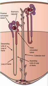
Image Showing Different Parts of nephrons: Image-credit-wikimedia commons
Bowman`s Capsule: The Bowman`s capsule was discovered by a British surgeon and anatomist Sir Willium Bowman in 1842. It is the dilated blind end of the nephron. It is a double walled structure around the glomerulus. The cellular layer which lies in close approximation with the glomerulus is known as the visceral layer and the outer cellular layer is known as parietal layer. The two layers are composed of single layer of flattened epithelial cells. The epithelial cells of the visceral layers are known as pedicels. The later are in close contact with the endothelial cells of the glomerular capillaries.
2. Renal Tubules: It is 3 cm long with a diameter of 60 µm, coiled structure arising from the base of the Bowman`s capsule and extending up to the collecting tubules. Anatomically, each renal tubule consists mainly of the following three distinct parts:
- The Proximal Convoluted Tubule
- The Loop of Henle and
- The Distal Convoluted Tubule
The Proximal Convoluted Tubule: It begins from the Bowman`s capsular space. It is a highly convoluted structure and about 14 mm long. It is lined by a single layer of brush border cuboidal cells. The cells have a numerous microvilli at their one side. The cells also have a large number of mitochondria in their cytoplasm. The water and solutes passed through the proximal convoluted tubule from the Bowman`s capsule.
The Loop of Henle: The proximal convoluted tubules lead into a U-shaped segment of renal tubule, called the loop of Henle after the name of its discoverer German physician Friedrich Gustov Jakob Henle. It is also known as nephron loop. The later enters straight into the medulla of the kidney, makes a hairpin bend and comes back to the cortex again. The major portion of the loop is lined by the flat epithelial cells.
The loop of Henle consists of two portions: first the descending limb of Henle then the ascending limb of Henle.
The Distal Convoluted Tubule: The loop of Henle is continued into the distal convoluted tubule. The length of the distal tubule is about 5 mm and is covered by columner epithelial cells. It empties its contents into the collecting tubule. Several collecting tubules from different nephrons join successfully and form the duct of Bellini which opens at the apex of the renal pyramid.
Functions of Different Parts of Nephron
Renal Corpuscle: It performs ultrafiltration of blood. In each minute 125 mm of excretory wastes, water and other substances are filtered from the blood in renal corpuscle and deposits as glomerulus filtrate in the Bowman`s capsule.
Proximal Convoluted Tubule: It performs filtration of Na+, Cl–, H2O. It reabsorbs HCO3–, K+, glucose, HPO4—, amino acid, uric acid, urea. It also does secretion of organic acid, base, H+, NH4+;
Loop of Henle: It performs reabsorption of H2O, Na+, Cl–, K+, Ca++
Distal convoluted tubule: It performs secretion of Na+, Cl–, HCO3–, K+, H2O, H+, NH4+,K+
Collecting duct: It does reabsorption of Na+, Cl–, H2O, K+, urea. It also does secretion of H+, NH4+, K+, etc.
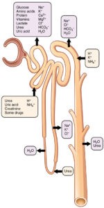
Image Showing Functions of Nephrons: Image credit-wikimedia commons
Functions of Kidney
Their basic functions include:
- Kidney filters out a variety of nitrogenous wastes products (urea, uric acid, creatinine, etc) and environmental toxins into the urine for excretion.
- Kidneys regulate the volume of extracellular fluid. In this case, It ensures the sufficient amount of plasma for maintaining the flowing of blood to fundamental organs.
- Kidneys help to control the osmolarity by making extracellular fluid more dilution or concentration.
- Kidneys help to regulate the maintenance of key ions such as sodium (Na+), potassium (K+), and calcium (Ca++).
- Kidneys help to prevent blood plasma too acidic or basic by regulating pH.
- Kidneys produce hormone like erytjropoietin which stimulates red blood cell synthesis. It also secretes rennin and antiotensin which help regulate blood pressure, salt, and water balance.
- Kidneys maintain gluconeogenesis. During prolong fasting, the kidneys synthesize glucose from amino acids and other precursors and release into blood. The kidneys are capable of supplying approximately 20% as much glucose as the liver at fasting.

