
Labeo rohita is the important food fish that belongs to the carp family Cyprinidae under order Cypriniformes of Class Actinopterygii. There are three crucial Indian major carp (IMC) such as, Labeo rohita, Catla catla and Cirrhinus cirrhosus, of which Labeo rohita is the most highly market-priced carp fish due to its attractive taste. It is also known as rui, rohu, or roho labeo. It can tolerate a wider temperature range, and it is the most adaptable and important cultured fish species in Bangladesh, India, Pakistan, and Myanmar.
Systematic Position
- Phylum: Chordata
- Sub phylum: Vertebrata
- Class: Actinopterygii
- Order: Cypriniformes
- Family: Cyprinidae
- Subfamily: Labeoninae
- Genus:Labeo
- Species:Labeo rohita Hamilton, 1822
Synonyms
Cyprinus rohita; Hamilton, 1822; Labeo rohita; Day, 1878; Shaw and Shebbeare, 1937; Labeo rohita; Bhuiyan, 1964
Fin Formula
D. 15-16 (3/12-13); P1. 16-17; P2. 9; A. 7(2/5). (Rahman, 2005)
Physical Description
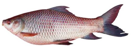
There is a proverb in Bengali that Rui (Rohu: Labeo rohita) is the king of fish, and Pui (vegetable) is the king of vegetables. So Rui is known to everyone as a very popular and delicious fish. The body of the rohu fish is spindle-shaped, and both sides of the body are symmetrical and flat. The head and tail are gradually narrower, but the head is 4-5 inches long. The dorsal part of the body is convex, but the surface of the head is more convex than the abdomen.
The snout is blunt, low, rarely swollen. The mouth is downward, and the lips are thick and fringed above and below the mouth, which is folded inwards. The mouth is located at the bottom region; the two corners of the mouth are curved backward, so the mouth is crescent-shaped. There is a pair of nostrils just in front of the eyes on the surface of the snout. The eyes are large in shape and have no eyelids. The cornea is transparent, which is covered by skin.
The whole body is covered with silver scales. The scales are smooth and arranged in rows. The upper lip of the mouth has a pair of barbells. The gill covers are wide. The surface scales of this fish are reddish, and the edges are black. This reddish color in the center of the scales becomes darker and brighter during the breeding season. Their dorsal and below the dorsal sides are brown, and their bellies are silvery white.
Their lateral line is complete, and there are 41-42 scales along this line. The dorsal fin has 15-16 soft rays, of which the first three rays are longer than the other rays. There are 16-17 soft rays in the pectoral fins, 9 in the pelvic fins, and 8 in the anal fins. The caudal fin is more bifurcated, and the caudal peduncle is short. They grow up to 200 cm in length and up to 45 kg in weight. They can live up to a maximum of 10 years. In Bangladesh, they are also called as Rohita, Ruhit, Rau, Nala, Garma, Naosi, etc.
Habit and Habitat
Apart from Bangladesh, this fish is found in North and Central India, Pakistan, Nepal, and Myanmar. They are usually river fish but also found in all freshwater bodies such as canals, beels, haors, baors(Lake), floodplains, ponds, streams, ditches, marches, etc. They are column feeders and mainly feed on plant-based food. Due to their slightly downward mouth with thick lips, they feed on aquatic plants, weeds, and occasionally take the rotten organic matter from the bottom region. When they are immature, they eat plankton. They also take fish meal, mustard oil cake, rice corn, etc. as supplementary food during cultivation in ponds.
Reproduction
They reach sexual maturity within two to three years. During the monsoon season, females and males take part in reproduction in flooded rivers, especially in aquatic vegetation areas. In one breeding season, a mother fish lays about two to thirty lac eggs, which can be more or less depending on the age, length, and weight. It also depends on the length and weight of the ovaries of fish. They lay their eggs in relatively shallow water and on the banks of rivers. Also, if artificial currents are created, rui fish can lay eggs in dams or ponds, without any worries. They usually lay eggs from June to August.
Embryonic Development of Labeo rohita
The diameter of fully swollen fertilized eggs is 4.1-4.8 mm, but their average diameter is 4.5 mm. The first cleavage occurs 45 minutes after fertilization. The second cleavage occurs within the next 5-6 minutes. The third cell division occurs within the next 15-20 minutes. The yolk invasion is half completed within 2 hours of fertilization. It reaches the Yolk plug stage in the next 1 hour.
Embryos can be identified 4 hours after fertilization. In the next 1 hour, the cluster of yolk becomes longer. A myotome is seen within the next 1 hour. In this stage, the head and tail part of the fetus is formed, the optic cup and 7-10 myotomes are also seen. Within 8 hours, 17 myotomes and capfer vesicles are seen, and the fetus begins to move. Eggs start hatching 14-18 hours after fertilization.

Figure: Embryonic development of Rui fish: Fertilized egg- (a) Newly formed bastodisk; (b) 2-cells phase; (c) 4-cells phase; (d) 8-cells phase; (f) 18-cells phase; (g) morula phase; (h) yolk plug phase, embryo; (i) elongation phase of yolk cluster; (j) About 2 hours before hatching.
Table: Different stages of larval life of Labeo rohita
| Hatching | Hatching time (hours) | |||||
|---|---|---|---|---|---|---|
| 0 | 6 | 12 | 24 | 36 | 48 | |
| Average total length (mm) |
3.78 |
4.70 |
5.31 |
5.49 |
5.85 |
6.20 |
|
Range (mm) |
3.62-3.83 |
4.62-4.78 |
5.22-5.40 |
5.25-5.84 |
5.71-5.99 |
6.07-6.33 |
|
Length of yolk sac (mm) |
2.52 |
2.94 |
3.15 |
2.79 |
2.52 |
2.90 |
|
Maximum height of yolk sac(mm) |
0.72 |
0.70 |
0.75 |
0.45 |
0.45 |
0.36 |
|
Body height along the dorsal fins (mm) |
0.90 |
1.00 |
1.08 |
0.90 |
0.90 |
0.90 |
|
Number of pre-anal myotome |
26 |
26 |
26 |
26 |
26 |
26 |
|
Number of post-anal myotome |
14 |
14 |
14 |
14 |
14 |
14 |
|
Eye diameter (mm) |
0.18 |
0.20 |
0.27 |
0.28 |
0.28 |
0.30 |
|
Eye color |
Light yellow |
Yellowish brown |
Black in the middle |
Black in the middle |
Dark black |
Black centered eye |
|
Length to the rear end of the notochord (mm) |
3.62 |
4.50 |
5.05 |
5.35 |
5.53 |
5.40 |
|
Direction of movement |
Irregular movement, rarely twisting up or staying sideways at the bottom. |
Irregular movement, comes from the bottom to the surface. |
Moves vertically, occasionally climbs up obliquely and does not take no rest. |
Performs vertical movements. |
Moves slowly with occasional jerking. |
Moves slowly with occasional jerking. |
|
Dorsal fin |
Absent |
Absent |
Absent |
Buds may be seen. |
Present |
Well-defined |
Larval Development of Labeo rohita
Hatchling
The yolk gradually becomes slender. Like the Mrigel and Catla, the embryos are similar in width from the posterior part to the yolk. The bulbous part of the anterior part of the yolk cluster is noticeable. The long slender part of the yolk cluster is smaller than Mrigel. Like other Puntius species, the heart is located in front of the yolk sac. A light pink marked posterior blunt part is seen on the dorsal part of the yolk. The eyes are slightly yellowish-brown.

Fig. Hatchling of Rui fish
6th Hours After Hatching
Dorsal fin buds are absent. The fetus moves irregularly. Rarely moves from the bottom to the top or rests on the bottom.
12th Hours After Hatching
Chromatophore can be seen only in the eye. The center of the eye to turn black due to the presence of chromatophore.

Fig. Larva of Labeo rohita: 12th hours after hatching
24th Hours After Hatching
Some black chromatophores are seen on the dorsal edge of the yolk sac. Gill arch becomes distinct. The tail looks like spatulate. There are a few black chromatophores on the head above the eyes. The color of the embryo is light yellow. The anus is clear. The auditory organ is completely distinct. The yolk sac is sharply attached to a sharped distal point. Various spots can be seen on the caudal fin. The notochord bends upwards. The pectoral fins do not have any fin rays.

Fig. Larva of Labeo rohita: 24th hours after hatching
36th Hours After Hatching
Lower lips become clear. The pectoral fins become apparent. A few black chromatophores are found on the whole dorsal edges of the yolk sac, the head and dorsal fins. No chromatophore is seen in the caudal region. The dorsal and ventral sides of the fetus are light yellow, but from the back of the tail region to the anus is dark black. The top of the notochord is bent upwards. The mouth exists as a tiny hole.

Fig. Larva of Labeo rohita: 36th hours after hatching
48th Hours After Hatching
The tip of the yolk sac is slightly convex like a catla, but it is not as straight as a mrigel carp. The embryos are yellow. The swimbladder is distinct, and the pectoral fins are noticeable. There is no chromatophore on the ventral side of the swimbladder. A row of black chromatophores can be seen from the posterior region on the right side of the auditory node to the base of the caudal fin.
The black chromatophores on the top of the head are apparent, and the chromatophores that spread to other parts of the body are large. Like the Mrigel fish, the pelvic embryonic fin folds begin at the front of the dorsal early fin folds. These fin folds do not contain any chromatophores. The gill arches are apparent, and the swimbladder is oval. The head is dark, and the body is slightly yellow.

Fig. Larva of Labeo rohita: 48th hours after hatching
72nd Hours After Hatching
The larval length is 6.95 mm. Its color is light yellow. The swim bladder is oval. Gill arch becomes specific. Some black chromatophores are seen on the head and the two eyes. The dorsal region to the anal area of the fetus is bright yellow. The tail region is yellow on the notochord.
You might also read: Mineral Requirements of Fish
Post larval development of Labeo rohita
96th Hours or 4 Day After Hatching
At this time, the length of the fish is 7.57 mm. Yellow chromatophores are seen in the head region behind the eye. The opercular part is distinct. The top of the notochord is curved. There are rows of distinct black chromatophores along the entire length of the lateral side of the embryo. Most larvae have an apparent reddish spot slightly above the anus. Yellow sac formation and caudal fin ray formation become completed. The two lips are of the same type but somewhat fimbriated type.
Under a magnifying glass, there is a crescent-shaped black region made of chromatophore in the tail region below the notochord. The chromatophores are densely embedded to form a straight line of a semicircle with a transparent area in the center. This semicircle is not continuous but apparent. Dorsal and pelvic fins folds exist.

Fig. Post larva of Labeo rohita: 96th hours or 4 days after hatching
5th Days After Hatching
At this time, the length of the fish is 8.5 mm. In most cases, prominent red spots are seen in the abdominal area. The swim bladder is oval. It is partially covered with black chromatophores. No chromatophores are seen in the folds of the dorsal and ventral embryonic fin folds. No rays can be seen in the folds of the dorsal fin. 5-7 rows of black chromatophores are seen on the body. The number of caudal fin rays is about 8-10. Black chromatophores are irregularly arranged in the caudal region.

Fig. Post larva of Labeo rohita: 5th days after hatching
6th Days After Hatching
The length of the fish is 10.5 mm. There are nine fin rays in the dorsal fin. Unclear rays can be seen in the anal fins, while buds can be seen on the pelvic fins. A few dark black chromatophores can be seen on the head. The dorsal side of the fetus is yellow. The swim bladder is divided into two parts. The anterior part of the fetus is round, and the posterior portion is long triangular. Black chromatophores cover both parts of the swim bladder.
The body has 7-8 rows of black chromatophores. The number of caudal fin rays is 22. On either side of the caudal fin, two black crescent-shaped bands are separated by a small pigment-free area. Black chromatophores can be seen below the notochord on the caudal peduncle. The caudal fins are slightly less forked than those of catla and mrigel fish. The ratio of the total length and the length of the base of the dorsal fin is 6.4: 1.

Fig. Post larva of Labeo rohita: 6th days after hatching
7th Days After Hatching
At this time, the length of the fish is 11 mm. Lips are thick. The top of the notochord is curved upwards. The ventral embryonic fin folds extend from the abdominal region to the anal fins while the dorsal embryonic fin fold is situated opposite to the anus. There is no pigment in the membranous junction area of the body. However, there are a few orange dots. There are two spots of black chromatophore on the caudal peduncle at the back of the origin of the fins ray. The caudal fin is less deeply forked than Catla and Mrigel carp, which contains 22 branched fin rays. There are few chromatophores at the base of the anal fins.
The pelvic fins have 2 or 3 fin rays with no chromatophore, while the dorsal fin contains 13 fin rays with no chromatophores at the margin of the dorsal fin. More chromatophores can be observed in the swim bladder region, but clear black chromatophores exist on the head. Black chromatophores are widespread throughout the body. However, the number of these chromatophores is not significant. The ratio between the total length of the body and the base of the dorsal fin is 6: 1.

Fig. Post larva of Labeo rohita: 7th days after hatching
8th Days After Hatching
At this time, the length of the fish is 12.5 mm. The dorsal fin has 14 rays, and a yellowish pigment is scattered near the base of those rays. No pigmentation can be noticed on the margins of the dorsal fins like Mrigel. The anal fin contains seven fin rays. A ventral embryonic fold is found in the anal region, which forms at the end of the abdominal area. The caudal fin provides 32 rays. The rays of the posterior part are branched. One-third of the base of these rays of this fin is orange. A few distinct black chromatophores can be noticed above the caudal peduncle. At the junction of caudal embryonic fin fold with the anal fin, a few black chromatophores and orange pigment spots are seen.
A few black and orange pigmented spots are also seen on the membranous anterior part of the caudal fin. These spots are similar to the catla but are more distinct than Mrigal. The body is golden yellow. Numerous black chromatophores are scattered throughout the body and head without any specific pattern, but it is few in the anal region. The ratio of the total length and the length of the base of the dorsal fin is 6.3: 1.
10th Days After Hatching
At this time, the length of the fish is 15.5 mm. From the thick upper lip hangs a fimbriated lower lip. There is a red mark from the anterior part of the abdominal region to the anus. There is no barbell. The color of the fish is golden yellow. The dorsal fin has 14 branched rays. Dark pigments cannot be noticed on the fins like catla. The anal fins have eight rays, and half of the base of the rays is covered with light pigment. The pelvic fin contains eight rays with no pigment. Like catla, a transparent fin fold is present that starts from the pelvic region to the anal region.
At the junction of the membranous caudal fin and the caudal peduncle is not covered by yellow pigment. There are 30 branched rays in the caudal fin. There are two light black crescent-shaped regions of the chromatophore on the caudal fin. These fins are covered by a band that extends to the tip of the posterior part of the origin. This crescent-shaped region on the caudal fin is not as clear as the catla. Such bands do not exist in Mrigal fish. The ratio of the total length and the length of the base of the dorsal fin is 5.7: 1.

Fig. Post larva of Labeo rohita: 10th days after hatching
12th Days After Hatching
At this time, the length of the fish is 19 mm. A pair of the maxillary barbell is distinct. The dorsal part of the body is colorful, and the ventral side is light yellow. 16 (3/13) rays can be seen in the caudal fin under the microscope. A yellow-orange pigment covers more than half the distance from the base of the dorsal fin. There are seven fin rays in the anal fin. The other fin rays are branched except the front one. The amount of black pigment is less on the half of the base of the fins rays than Mrigel, but it has the same as catla. The black chromatophores of the dorsal half of the body are more pronounced. The pelvic fin contains seven fin rays but no fin ray on the membranous anterior margin of the caudal fin. There are more yellow pigments on the dorsal side.
The two chromatophores are seen to merge a little behind the origin of the fins. This junction area is dark in color but not precisely crescent-shaped like catla. Orange pigments can also be noticed in this region. A wide triangular band can be observed in front of the origin of the rays on the caudal peduncle. In most cases, the band extends along the entire width of the front of the head. These chromatophores are present under the microscope, but they do not co-exist. The caudal fin contains 34 fin rays. Orange pigments are scattered in the first half of the base of these rays. The ratio of the total length and the length of the dorsal fin is 6.3: 1.
15th Days After Hatching
At this time, the larvae are 23 mm in length. There is a hanging lip with a pair of barbells. Orange pigmented spots exist on yellow pigment. Moreover, such orange spots exist among the rays of the dorsal fin. The anal fin contains 7 (2/5) fin rays. Only one-third of the base of the rays has orange pigment. Anus exists in front of the anal fins like Mrigel. However, this situation is not seen in catla.
There are seven rays in the pelvic fins. The black pigment can be seen on half of the base of the pelvic fin. There is a clear band of black chromatophores along the entire length fin ray in front of the caudal peduncle. The origin of the caudal fin ray is divided into two halves, and the caudal fin extends posteriorly. The number of caudal fin rays is 34. There are six unbranched fin rays on each side of the caudal fin. The ratio of the total length and the length of the base of the dorsal fin is 5.4: 1.

Fig. Post larva of Labeo rohita: 15th days after hatching
18th Days After Hatching
At this time, the length of the fish is 25 mm. Black chromatophores cover the body. Such chromatophores are more abundant above the lateral line. The body color on the lateral line is light yellow. On the other hand, the lower part of the lateral line is yellowish-white. The body is covered with scales. However, the scales of the abdominal region are not clear, especially near the caudal peduncle and the abdominal region.
When the length of the fish becomes 24 mm, the first scales are observed at this time. At this stage, 4-5 rows of scales exist next to the opercular margin to the beginning of the dorsal fin. At this time, a pair of barbells also exist. Pelvic fin contains nine fin rays. The half of the base of this fin rays contain orange pigment. Two distinct gray half-crescent-shaped structures are present on each lobe of the caudal fin.
20th Days After Hatching
At this time, the length of the fish is 26 mm. There is a pair of barbells on the margin of the thick hanging lips. Each barbell originates from a notch in the mandibular region. The body is completely covered the golden-colored scales. The color of the particular part of the body above the lateral line is yellow. The color of the underside of the lateral line is light dirty yellow. The transparent black chromatophores are spread all over the body. There is a dark-colored band above and below the lateral line, which runs from the next gill cover to the caudal region.
Several black chromatophores exist along the margin of the dorsal fin. The rays of the pelvic fins are colorless and not transparent. The posterior border of the band is slightly concave, but the front edge is irregular. Two spots separate this dark band, and two light crescent-shaped areas are seen. Crescent-shaped areas contain a small black spot. This spot also exists in an outer part of the dark caudal fin band. Orange pigment covers the caudal fin rays with a few spots at the distal ends. The caudal fin contains 34 rays, the rays of the margin of each side are unbranched.
25th Days After Hatching
At this time, the length of the fish is 30 mm. Their color is light brown with a tan hue from operculum. The barbell is noticeable. The upper margin around the eyes is slightly orange. The scales are distinct. The dorsal fins are slightly orange with small black. The base of this fin is yellow. The pelvic fin contains 7 (2/5) fin rays. Except for the one-third part of the first few rays, the other rays show orange pigment.
The rays on the back of the pectoral fins contain no pigment. The first few rays contain black and orange pigments. There is no pigment in the pelvic fins except for the second branched fin ray. The dark band in the caudal region is fully visible. Under the microscope, the band appears round. Large black chromatophores cover this band entirely. The more distinct pigment can be seen in the pelvic fins. The upper part of the caudal fin is larger and pointed, but the base is slightly smaller, and the margins are rounded.
Table: Difference among Rui, Catla and Mrigal during the development stages
| Mrigal (Cirrhinus chirrhosus) | Catla (Catla catla) | Rui (Labeo rohita) |
|---|---|---|
|
Hatchling: The thinner part of the yolk is more significant in length than the thicker part. Yolk is more or less club-shaped. Most hatchlings have 28 prenatal and 14 post anal myotomes. |
The thick and thin part of the yolk is equal in length. In most cases, there are 26 prenatal and 14 post-anal myotomes. |
Like catla. |
|
24 hours after hatching: The narrow end of the yolk sac did not end at a pointed end. |
Like mrigal |
The narrow edge of the yolk sac ends in a slightly pointed tip. |
|
36 hours after hatching:The anterior profile of the yolk sac is more or less straight. |
Anterior profile yolk sac is convex. |
Like catla |
|
48 hours after hatching:The anterior profile of the yolk sac is more or less straight. |
Anterior profile yolk sac is convex. |
Like catla |
|
72 hours after hatching: There are a few black chromatophores in the caudal region. There is no reddish impression in the operculum region. |
The opercular region has a reddish tinge. There are dark and somewhat triangular spots on the caudal peduncle. |
The caudal region has a few light pigmented spots behind the notochord. There is no reddish tinge in the opercular area. |
|
96 hours after hatching: The dorsal fins have black chromatophores at the front of the dorsal fin buds. Black chromatophores exist below the apex of the notochord. These chromatophores are confined to a semi-circular area. The reddish color is seen in the abdominal region. Such characters are not seen in Rui and catla. Lips thin. The dorsal fin is separated from the embryonic fold. |
A semicircular mark with black chromatophores is seen on the ventarl side of the caudal fin just in front of the edge of the notochord. The arc of the semicircle is not clear, and the straight line of the arch is somewhat concave. The margin of the lips is thick. The abdomen above the anus has a reddish tinge. The dorsal fin is separated from the embryonic fold. |
The black chromatophores lined beneath the notochord form a crescent-shaped semicircular region. In most species, a red spot is seen on the bare eye just above the anus at the beginning of the pelvic fins fold. The edges of the lips are slightly fimbriated. |
|
Five days after hatching: The number of caudal fin rays is 7. They are not branched. There are no rays in the front part of the dorsal fin. As a result, it separates from the embryonic fold. |
At the bottom of the notochord, 18 rays are visible, with branches at their distal ends. On the front of the dorsal fin, there are six rays which are different from the embryonic folds. |
About 8-10 fin rays are seen in the caudal fin. They are branched. In most cases, the abdomen is clearly reddish. At this stage, dorsal fins are seen, which are different from embryonic folds. |
|
Six days after hatching: The dorsal half of the body is yellowish-green. There are crescent-shaped two light marks on the caudal peduncle. Anal fins exist. |
The body is light yellow. The caudal region has two crescent-shaped regions with chromatophores. Anal fin absent. |
The dorsal part of the body is yellow. The two crescent-shaped parts made of chromatophore on the two lobes of the caudal fin, which are separated by a colorless area. Anal bud exists. |
|
Seven days after hatching: On the caudal peduncle, a triangular structure formed by black chromatophore that can be seen in front of the origin of the caudal fin ray. The caudal fin is not as deeply forked as the catla. The dorsal fin is completely separate from the embryonic fin fold. From the front of the membranous part of the caudal fin to the beginning of the caudal fin ray, a clear yellow pigment is spread. |
Two small crescent-shaped structures with black chromatophores are present just behind the beginning of the caudal fin. The caudal fins are more forked than Mrigel and Rui. The dorsal fin is completely separated from the embryonic fold. There is no yellow pigment from the front of the membranous part of the caudal fin to the beginning of the caudal fin ray. |
Two clusters of black chromatophores exist on the caudal peduncle. The caudal fin is less forked than catla. The dorsal fin is completely separated from the embryonic fold. There are several spots of orange pigment from the front of the membranous part of the caudal fin to the beginning of the caudal fin ray. |
|
10 days after hatching:Lips thin, like full-grown adults. There is no black chromatophore at the margin of the dorsal fin. A triangular area with black chromatophores can be seen just in front of the origin of the caudal fin. |
Lips thick. The mouth is slightly upward. The margin of the dorsal fin is dark blue due to chromatophore. Somewhat anterior to the origin of the fin ray, there are two distinct chromatophores on the caudal peduncle. A triangular region with black chromatophores is present below the notochord. This region is less clear than Mrigel. |
The thick fimbriated upper jaw is present, which hangs over the lower jaw. There are no chromatophores on the margin of the dorsal fin. Two faint black chromatophores exist on the caudal fin ray. Beneath the sharply curved notochord, there is a triangular region with a more or less broad base formed by black chromatophores. Each chromatophore is clear. |
|
15 days after hatching: The dorsal half on the body, especially on the lateral line, is richer in pigment than the rui and catla. As a result, a thin dark parallel line exists on the yellow tinge. There is no crescent-shaped pigment in the tail region. Scales can be seen. |
The margins of the dorsal fin are black. Two-third part of the dorsal fin ray from the base is covered with light yellow pigment. Black chromatophores cover the triangular-shaped region on the caudal peduncle. Scales are not seen. |
Thick hanging lips. Two barbells can be seen. Two-third of the base of the dorsal fin is orange pigmented. The caudal fin region contains black chromatophores that lengthen posteriorly, which divide the caudal fin into two lobes. Scales are not seen. |
Economic Importance
It has a high demand in the market due to its attractive taste. Besides, its nutritional value is very high. For every 100 grams of rui fish contain 16.4 grams of protein, 1.4 grams of fat, 680 mg calcium (Ca), and 223 mg phosphorus(P). In Bangladesh, Rui, Catla, and Mrigel fish account for 22.5% of the total fish production.
Reference
Chakraborty, R.D. and Murty, R.S.V. 1972. Life history of Indian major carps, Cirrhina mrigala (Hamilton), Catla catla (Hamilton), and Labeo rohita(Hamilton). Journal of Indian Fisheries Society of India. 4:132-161.
Kabir, A.K.M. N. and Mia, Mohiuddin. 2018. Fisheries Biology(in Bengali). 2nd edition. ATM Publications, 38/3, Banglabazar, Dhaka. pp. 623.
Rahman A. K. A. 2005. Freshwater Fishes of Bangladesh. 2nd edition. Zool. Soc. Bangladesh. pp. 394.
Shafi, M. and Quddus, M. M. A. 2003. Bangladeshher Matshya Sampad (in Bangali). Kabir Publications, 38/3, Banglabazar, Dhaka. pp. 345.