Scales are small plate-shaped dermal or epidermal structures that are found in the outer skeletons of fish, reptiles, or some mammals. The skeletons of many vertebrates are covered by two types of scales, namely epidermal and dermal. Epidermal scales originate from the malpighian layer of the epidermis. Such scales are found in terrestrial vertebrates such as reptiles, birds and mammals. Afterwards, dermal scales emerge from the mesenchyme of fish. Such scales are made up of small, thin, thorny and crushed or bony plates that stick closely to each other. The outer skeleton of a fish is called scales.
The body of an ideal fish is covered by thin scales. The scales develop as external growths of the epidermis or skin. The epidermis contains numerous mucus cells. These cells secrete mucus or slime, which prevents parasites, fungi, pathogens, etc. from entering the skin easily. Most fish bear scales. Agnatha and catfish have no scales. Some fish, especially paddle fish (Polyodon), mirror carp (Cyprinus carpio) have partial scales. Other fish such as trout and freshwater eel have very small scales. Scales cover most of the body and protect the skin from injury.
The scales contain a variety of pigments that give the fish a variety of colors. The scales form a lateral line in the body of the fish along the side of the body and play an important role in detecting vibrations in the water as it acts as a sensory receptor.
When the fish hatches from the egg, its body is covered by small scales. As the fish grows so does the scales. However, the number of scales remains the same throughout life but the lost scales can be restored at some point. A small circular growth ring is formed in the scales and this ring is called circuli or circulus (in singular). Circulus formed in summer are quite wide whereas circulus formed in winter are intertwined. In that densely enclosed region a black circle is formed which is known as the annulus. The age of the fish is determined by counting the number of annulus in scales.
Types of Scales
Fish scales can be divided into four based on shape, namely:
- Placoid
- Cycloid
- Ganoid
- Ctneoid
Fish scales can also be divided into two based on their structure, namely:
- Placoid
- Non-placoid
There are three types of non-placid scales, namely:
- Cosmoid
- Ganoid and
- Bony Ridge
Bony ridge scales are divided into cycloid and ctenoid based on the thorns. The following is a description of the different types of scales:

Placoid Scales
Such scales are found only in cartilaginous fish (Chondrichthyes) but are absent in the subclass Holocephali. The scales cover the skin like sand grains. Placoid scales are arranged in different rows individually to form the outer skeleton. At the bottom of each scales is a base plate and a pointed thorn arising from the base. This thorn is curved backwards. As a result, the scales protect the skin from abrasive injuries. Each base plate is made up of calcium-rich tissues. Thorny structures are inserted into the dermis with the help of sharp`s fibers and other fibers. The fork is made up of dentin with an enamel coating on the outside.
Development of Placid Scales
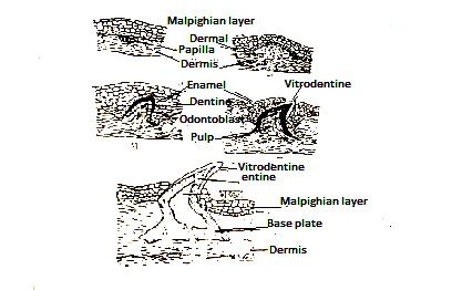
The development of a placoid scales begins with an increase in the number of dermal cells accumulated at different points in the stratum germinativam (Malpighian layer). These dermal cells form a papilla that grows upwards and pushes the Malpighian layer to form an arch-like structure. In this region, the Malpighian layer cells become columnar and glandular to form enamel organs. Dermal papilla, roughly a base plate and a thorn-like papillary surface, called odontoblast, form a distinct layer.
These cells also cover the papilla by secreting dentin on the outer surface. The surface of the central cells of the papilla does not cover the papilla. Papillary cells do not exist and do not form pulp. The thorns of the scales slowly push the surface upwards. The cells in the enamel organ also get known as amyloblasts before they rupture. These cells secrete a layer called vitrodentin to form a coating around the spine.

Development of Placoid scale
The mesenchyme cells of the dermis, which surround the dermal papilla, form a fibrous base plate by secreting a substance resembling tooth cement. Thus a placoid scales become partially dermal and partial epidermal from the basal side. The thorns protrude towards the top of the skin while the base plate is hidden in the dermis.Placoid scales become larger and change to form shark teeth. In vertebrates, teeth erupt in the same way and have a comparable structure. Therefore, placid scales and vertebrates teeth are also considered to be coexistent.
You might also read: Tilapia: Physical Description, Habitat, Reproduction and Economic Importance
Cosmoid Scales
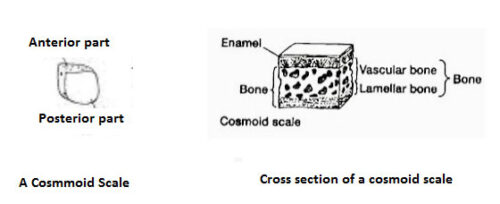
No such live fish can be found. The scales were present in the bodies of some Ostracoderms, placoderms, and extinct sarcopterygian. There are four levels in this scale. On the outside of the scales is an enamel-like thin and hard vitrodentin layer, below this layer is a hard and non-cellular cosmin layer, on the inside of the cosmic layer is the isopedine layer, which consists of the duct and the ossicular layer. The growth of these scales occurs towards the edge. It does not grow here because there are no living cells at the bottom. In the case of lung fish, the original cosmoid structure has changed to cycloid scales.

Ganoid Scales
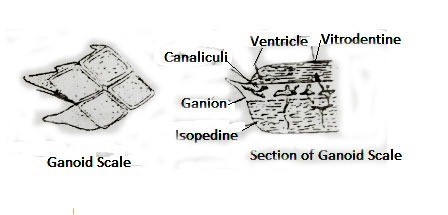
Ganoid scales consist of thick, usually diamond-shaped plates. The roof also has tile-like scales attached side by side to form a bony covering. In some cases, the scales overlap. Such scales are found in Chondrostian (Polypterus, Acipenser) and Holostian (Lepisosteus) fish. One of these fish is called ganoid fish. Pelioniscoid ganoid scales exist in the bichir (Polypterus). These scales consist of an enamel-like ganoid layer on the outside, a dentin-like cosmin layer on the middle, and a bony isopodine layer on the inside. Lepidostoid ganoid scales are present in Lepisosteus. The scales have ganoin on the outside and isopedine on the inside. This scale increases in all directions.

Bony Ridge
Such scales are thin, piercing. It does not have enameloid and dentinal layer. This type of scales is found in most living bony fish(Osteichthyes). It is of two types, namely:
- Cycloid and
- Ctenoid Scale
Cycloid Scales
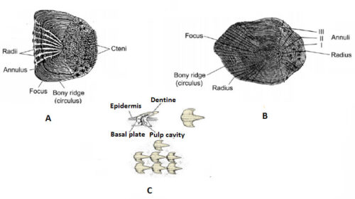
Such scales are found in lungfish, some Holosteans and non-teleostean such as carp (Cypriniformes), hilsa (Clupeiformes) and cod (Gadiformes). Such scales consist of round plates. The center of this scales is called the focus. Many concentric circular growth lines can be seen from the focus. The upper part of these growth lines is made up of thin bony and the lower part is made up of fibrous connective tissue.
At certain times of the year, especially in winter, the growth of fish decreases as the growth of scales decreases, resulting in the formation of ridge or circuli in the scales. The large circulus is known as annulus which is formed annually. In some species, many radiuses are seen near the focus. The scales overlap each other. The scales are located in a small pocket on the front of the dermis and the rear part is exposed.
Ctenoid Scales
Such scales can be seen in modern advanced teleostean fish such as perch (Perciformes), sunfish, etc. The shape, texture and decoration are exactly like cycloid scales. However, there are small thorns(cteni) in the open back side. This is why this type of scales is called ctenoid scales. These scales are more firmly attached.
Some fish such as Jew fish (Johnius) flat fish (Cynoglossus), flounder fish have both cycloid and ctenoid types. These fish have ctenoid scales on the surface and cycloid scales on the numerical side.

A. Ctenoid scale B. Cycloid scale and C. Placoid scale
Modification of Scales
Different types of modifications are observed in fish scales. Some fish such as electric fish, catfish (Siluriformes), eel (Anguiliformes) have small scales that are placed in the dermis. Some fish have scales transformed into different organs. Shark jaw teeth, dorsal spines of dogfish (Squalus) and chimerid (Chimera), tail spines of stingrays (Dasyatidae), saw teeth of saw sharks (Pristis), gill racker of basking sharks (Cetorhinus), etc are conversion of placoid scales.
The lancet of the sergeon fish (Acanthurus) is formed by converting the tinoid scales. The abdominal scutes of the herring (Clupeidae) are transformed into cycloid scales. Lepidotricia of the fins and dermal bones are the modifications of bony ridge. Many bony fish scales have been transformed to form a hard groove for the lateral line organ as well. Bone plates or scutes of stargeon fish (Acipenser) are also the modification of scales . Multiple transformations of scales have occurred due to the urge to live, especially due to self-defense, food intake, etc.
Uses and Functions of Fish Scales
Scales are protective external skeletons and have multiple functions and uses. The functions and uses of scales are mentioned below:
1. Fish classification: The importance of scales in fish classification is immense. The number of scales varies from species to species. Along the lateral line of the fish, rows of scales above and below it are used to identify family, genera, and species in hierarchy.
2. Life history of fish: With the growth of fish throughout life, scales also increase. As a result of growth, some concentric circular lines are seen on the scales which are called growth lines. These lines are also created to vary the physical growth of the fish in different seasons. During the winter, the growth of fish in the region is hampered in winter, resulting in the formation of a large line every year, known as the annulus. From these lines fish breeding, seasonal growth, annual growth etc. are known. In addition, Atlantic salmon scales have a spawning mark that can be used to determine how many times a fish has spawn. During reproduction, there is a sudden change in the existing circulus in the scales.
3. In self-defense and food hunting: The thorn that is formed by the transformation of scales plays an important role in fish self-defense and food hunting. In addition, the scales protect the fish from parasites as a protective coating.
4. Gender selection: In the breeding season, some species of matured male fish have different colors (Punteus).
5. Adaptation: Different types of fish scales and their color help the fish to adapt in different environments.
6. Locomotion: Some fish scales (Anabas) help the fish to move.
7. Osmosis: As the water is impermeable, water from the reservoir helps in controlling the infiltration by entering the body of the fish or discharging water from the body into the reservoir.
Difference Between Placoid scale and Cycloid scale
|
Placoid scale |
Cycloid scale |
|---|---|
|
These scales are plate shaped. |
These scales are round and circular in shape |
|
This scales are found in the skin of cartilaginous fish. |
Cycloid scales are found in soft-feathered hardy fish such as Labeo, Catla, Punteus, etc. |
|
Such scales are called dermal denticles. |
This is called bony ridge. |
|
Such scales have disc or disc-shaped base plates. |
Such scales have no base plate. |
|
These scales originate from the epidermis and dermis. |
These scales originate from the dermis. |
|
Such scales have a thorn that is exposed in the epidermis. |
Such scales do not have any thorns. |
|
Such scales do not have circular, and chromatophore. |
Such scales contain numerous circulus, radii and chromatophores. |
|
The scales contain an enamel-like hard and transparent material called vitrodentine |
The scales do not contain hard and transparent ingredients like enamel called vitrodentin. |
|
It does not have a growth line. |
There are growth lines. |
|
It is transformed into a bone-like structure, especially the dorsal fins of the Squalus, the tail thorns in the stingrays (Dasyatidae), and the saw-like teeth in the saw sharks. |
It is transformed into abdominal squat (Clupeidae) |
