The tissue which are responsible for locomotion and for the movement of the different body parts is called muscular tissue. The muscle cells are often called muscle fibres due to elongated shapes of the cells. Muscle tissue is a specialized tissue which composed of fibres of muscle cells. They control the movements of an organisms by applying forces to different parts of the body.
Muscle tissue is an elongated tissue which can range from several mm to about 10 cm in length and from 10 to 100 µm in width. Muscles contain contractile protein with contraction and relaxation characteristics which is responsible for movement. Skeletal muscle tissue contains elongated muscle cells, known as muscle fibres responsible for the movement of the body. There are various types of muscle tissues in the body. The mammalian body contains three types of muscles tissue: These are skeletal or striated muscle tissue, smooth muscle or non-striated muscle and cardiac muscle.
Properties of Muscular Tissue
- The muscle cells have the ability to contract forcefully. In this case, muscles can only pull but never push.
- The muscles have the ability to shrinking or bounce back to the muscle’s original length after being stretched.
- The muscular tissue reacts to a stimulus which may be delivered from a motor nerve or hormone.
- A muscles have the capability to be stretched.
Classification of Muscular Tissue
According to distribution, structure and function, the muscular tissues are of three types:
- Based on distribution: Skeletal, visceral and cardiac;
- Based on structure: Striated and non-striated;
- Based on function: Voluntary and involuntary.
1. Striated Muscle or Skeletal or Voluntary Muscle
In the fresh state human striated muscle looks pink color partly due to presence of myoglobin, the pigment in the muscle fibres and partly due to the rich vascularity of the tissue.
Fig. Showing different Skeletal Muscles
Structure
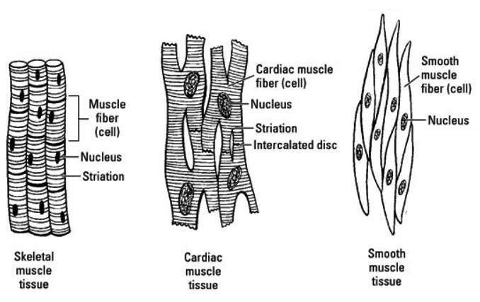
The shape of a muscle cell (fibre) of skeletal muscle is long, cylindrical, multinuclear, the ends tapering to a point. It extends in parallel direction with longitudinal length of muscle from point of origin to point of insertion(attached place with bone) often through aponeursis , the extended tendons.
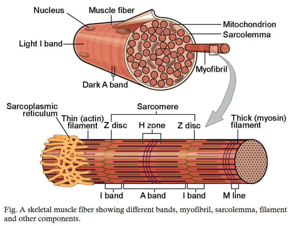
The length and breadth of the muscle fibre vary from 1.0 to 40 mm and 0.01 mm(10µm) to 0.1 mm(100 µm) respectively. Each muscle cell contains its own thin, tough and transparent covering called the sarcolemma as well as a thin connective tissue sheath called endomysium that remains just outside the sarcolemma. A bundle of several muscle fibres is wrapped by a connective tissue sheath called perimysium. Several such bundles, each consisting of few muscle fibres are grouped together to form the structure of muscle. The muscle as a whole is enclosed by a tough connective sheath called the epimysium. Each muscle fibre is filled up with cytoplasm known as sarcoplasm.
Inside the sarcolemma, elongated multiple nuclei and transversely striated myofibril are embedded in the sarcoplasm. The sarcoplasm contains other constituents like mitochondria (sarcosomes), golgi bodies, and around the myofibril sarcoplasmic reticulum are present.
Myofibril
The structural peculiarities of skeletal muscle are the alternate light and dark bands or shades (transverse striation) and thick longitudinal strands. The dark band is anisotropic and the light band is isotropic when seen with polarized light. Hence, the dark band is called as A-band and light band is called as I-band.
The I-band is intersected by a thin dark line, the Z-line(Double`s line or Krause`s membrane). The amount of substance included between two successive Z-lines of a myofibrils is considered as contractile unit and is known sarcomere. Other bands in the myofibril are small H-zone( Hensen`s line) bisecting A-band. The myofibril composed of mainly two types of protein filaments. The thick filament is known as myosin filament and the thin filament is known as actin filament.
Distribution
Striated muscle is called skeletal muscle as it is generally attached to the bone of the skeleton. In addition, muscles are also distributed in the tongue, upper part of the oesophagus, etc.
Function
As the skeletal muscle is directly under the control of the will so it is also called voluntary muscle. It is concentrated in body movement and in locomotion. Skeletal muscle is supplied with somatic (voluntary) nervous system.
2.Non-striated muscle or Smooth muscle or Visceral muscle or Involuntary muscle.
Structure
The shape of smooth muscle fibres are elongated spindle-shaped cell with fine tapering ends. The average length of the muscle fibre is about 20 µm and the width is about 6 µm at the central widest portion. The sarcoplasm contains fine myofibrils which are lack of striation (cross striation). Smooth muscle fibres do not have distinct sarcoplasm. The single nucleus is elongated, oval or cylindrical in shape and is placed at the center of the muscle fibre.
Distribution
It is mainly present in the visceral organs. It forms the main part of the digestive tract from the centre of the oesophagus to the anus. It is also present in ducts of glands, blood vessels, respiratory, urinogenital and lymphatic systems of the body.
Function
The non-striated visceral muscles are involuntary and cannot be moved at will. Almost all systemic functions done by the different visceral organs are controlled to some extent by the smooth muscle present in those organs.
These types of muscle fibres are supplied with automatic nerve fibres (systematic and parasympathetic). These nerve fibres control the function of the muscle.
3. Cardiac Muscle
Cardiac muscle is involuntary but striated and contracts rhythmically and automatically.
Structure
Cardiac muscle is made up of muscle cells. These are of 2-22 µm in diameter. Under light microscope, the cardiac muscles appear as syncytium (cytoplasmic continuation between neighboring muscle fibres).However, now it has been revealed by electron microscope studies that heart or cardiac muscle is made up of distinct individual cells.
The heart muscles look like syncytium because adjacent cell membranes are in very close contact with each other through special structures called intercalated discs. These discs appear at the dark lines which pass in irregular step-like configuration across the fibre at fairly regular interval. Each cell contains centrally placed single oval nucleus, sarcoplasm and myofibrils.
Distribution: Heart is made up of this type of muscle.
Function: The contraction and relaxation of the heart (cardiac) muscle is not under our will hence it is involuntary muscle.
Table: Showing comparison of three muscular tissue
|
Muscle types |
Location |
Appearance |
Function |
Nature |
|---|---|---|---|---|
|
Skeletal muscle |
It is present in Skeleton |
Striated, multinucleated, Fibres are parallel |
Movement, heat, posture, etc. |
Voluntary |
|
Smooth muscle |
It is found in gastrointestinal tract, uterus, eye, blood vessels. |
Striation is absent, one central nucleus is present |
Peristalsis, blood pressure, pupil size control, erect hairs |
Involuntary |
|
Cardiac muscle |
It is present in Heart |
It is striated with one central nucleus |
Pumping blood in the body continuously. |
Involuntary |
Functions of Muscular Tissue
- Skeletal muscles helps to provide rigidity to our body for maintaining our body movements such as walking, chewing, running, lifting, etc.
- Muscle system produce a continuous contractile power that help to maintain an erect or seated position or posture.
- Muscular system helps in respiration. In this case, muscle fibres spontaneously drive movement of air into and out of our body.
- Muscle tissue can generate heat during contraction of muscles which is required for the upkeep of temperature homeostasis.
- Smooth muscles help to supply nutrients through digestive tract. It also helps to passes out urine from the body.
- The smooth muscles helps to enhance secretion from the glands through contraction of muscles.
- Muscle fibres regulate blood pressure through contraction or relaxation of blood vessels.
- It also helps to do distribution of blood throughout the body.
- Muscle tissues help in the pumping blood. In this case, the heart receives blood continuously and release it to all the tissues of the body and organs.
- In many cases, muscle tissues help to protect many delicate internal organs through proper covering them.
- It also maintains the integrity of the body cavities.


