
The cell is the fundamental structural, biological and functional unit of living things. The typical cell contains the most outstanding visual and functional feature, the nucleus. The nucleus is the most prominent organelle which occupies about 10 % of the volume of the cell. The nucleus is the double membrane-bounded dense round cellular component which contains a genetic material DNA in chromosome and generally, it is located at the center of the cell.
Microbiologist Antonie van Leeuwenhoek first observed the nucleus in the red blood cells of salmon fish while in 1804, Franz Bauer also described the nucleus but Scottish Botanist Robert Brown (1831) observed the dense circular region inside the plant cell and he called it nucleus and described more details about the nucleus.
The nucleus is the biggest cell organelle found in the cytoplasm of all eukaryotic cells of plants and animals. The size of the nucleus varies from cell to cell. The size is directly proportional to that of cytoplasm. The average size of the nucleus in mammalian cells is approximately 6 µm in diameter. The mammalian matured erythrocytes and matured sieve tubes of higher plants contain no nucleus. The contents of the nucleus are present in the nucleoplasm which is similar to the cytoplasm.
The shape of the nucleus normally remains related to the shape of the cell. The shape of the nucleus also varies which may be circular, oval, disc-shaped, elongated, lobed in WBC (white blood cell), C-shaped in Vorticella, pyriform in Paramecium, moniliform in Spirostomum, spindle or elliptical in sperm.
The eukaryotic cells usually have a single nucleus (mono-nucleated cell), sometimes, the cell may contain two nuclei (bi-nucleated cell) and the osteoclasts bear many nuclei (poly-nucleated cell) while the mammalian red blood cells (RBC) have no nuclei.

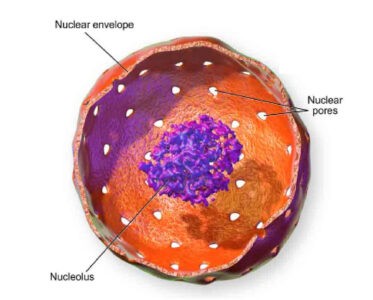
Structure of Nucleus
Structure of Nucleus
The nucleus consists of the following structure:
- The nuclear membrane
- The Nucleoplasmt
- The Nucleolus and
- Chromatin Fibers
The Nuclear Membrane
Nucleus is covered by the nuclear membrane. It is made up of inner and outer membrane. This membrane makes separation the cytoplasm from the nucleoplasm. It is also known as nuclear envelope, nucleolemma or karyotheka. It is derived from the membrane of the endoplasmic reticulum.
Each membrane is 75-90 Å thick and is made up of lipoprotein. Two layers (outer and inner membrane) are mainly separated by a fluid-filled space of 100-150 Å which is also known as perinuclear cisternae. The outer layer of the membrane is attached to the (ER) endoplasmic reticulum at several points. Ribosomes remain attached to the outer surface of the outer membrane and hence it looks rough. At certain points, the nuclear envelope is interrupted by the structures called pores. Both membranes are in continuity around the margins of these pores. Nuclear pores act as selective diffusion barriers between the nucleus and cytoplasm.
Nuclear Pores: The apertures (50-80 µm in diameter) present in the nuclear envelope are known as pores, within each pore octagonal shaped electron dense ring is present which is called annulus (60 nm in diameter). It consists of eight granules that present at both the nuclear and cytoplasmic surface. The pores and annulus together are referred to as the pore complexes.
Functions of Nuclear Membrane
- The nuclear membrane makes separation the contents of the nucleus from the cytoplasm.
- It allows the free exchange of ions between nucleoplasm and cytoplasm.
- It protects the internal structure of the nucleus.
- It attaches the structural elements of the cytoplasm and nuclear components.
- Ribosome remains attached on the outer surface of the membrane helps to synthesize protein.
- Golgi bodies and Endoplasmic reticulum (ER) are formed from the nuclear membrane.
- It consists of phospholipids that help to form a lipid bilayer.
- It helps to regulate the shape of the nucleus.
- It provides assistance to regulate the flow of molecules between the cytoplasm and nucleoplasm through nuclear pores.
Nucleoplasm
Nucleoplasm is a relatively clear granulated semi-liquid, slightly acidic gelatinous substance which is present in the space between the nuclear membrane and nucleolus. This semi-aqueous material is similar to the cytoplasm. It is also called nuclear sap or karyolymph. It contains a thread-like elongated structure which is known as chromosomes.
It also contains different types of chemical substances such as:
- Dissolved phosphorus
- Dissolved Salts
- Ribose sugar
- Phosphoproteins
- Nucleotides
- Nucleic acids
- Different enzymes
- The trace amount of minerals
- Water
Functions of Nucleoplasm
- Nucleoplasm contains the nucleolus and chromosomes.
- It helps to protect the contents of the nucleus.
- It support the nucleus by helping to maintain its shape.
- It provides a medium by which different materials like enzymes, nucleotides can move throughout the nucleus.
Nucleolus
The nucleolus is a discrete densely stained membrane-less spherical body which is found in the nucleoplasm of the nucleus. It is composed of RNA and proteins. It is attached to the special regions of particular chromosomes, known as nucleolar organizer regions which help to synthesis ribosome by transcribing and assembling ribosomal RNA subunits.
During the karyokinesis or first phase of cell division, it disappears completely and again reappears after cell division. It is composed of a large amount of ribosomal protein and ribosomal RNA (rRNA).
Structurally, it is made up of four zones:
1. Granular Zone: It is the peripheral part of the nucleolus which contains granules of ribonucleoprotein.
2. Fibrillar Zone: It is composed of fibrils of ribonucleoprotein.
3. Amorphous Zone: It has low electron density and it is found only in a certain nucleolus.
4. Chromatic Zone: Surrounding the nucleolus, a shell-like structure is present which is known as perinuclear chromatin. This may be in the form of a thick continuous wall as an exocrine pancreatic cell. From perinuclear chromatin, the intranuclear chromatins arise.
Functions of Nucleolus
- It is the most active site for synthesis of RNA.
- It helps in the formation of ribosomes.
- It plays an important role in the synthesis of protein by producing ribosomes
Chromatin Fibers
They are elongated thread-like coiled structures within the nucleoplasm of the nucleolus. It is also known as nuclear reticulum. They got the name chromatin (Gr. Chroma=color) due to their colorful nature during cell staining when it is viewed under microscope. They are twisted, fine anastomosed chromatin fibers and are uniformly distributed in the nucleoplasm. These chromatin fibers are observed only during a resting state of cell division. During cell division, chromatin fibers become thick ribbon-like structures, called chromosomes. Chromatin fibers are of two types: Euchromatin and Heterochromatin. Actually, chromatins are composed of histone protein, DNA and trace amount of RNA. In this case, DNA and histone protein exist in chromatin in 1:1 ratio.
Functions of Chromatin Fibers
- They bear hereditary instructions.
- They regulate the different cellular process.
General Functions of Nucleus
- The nucleus controls nutritive, respiratory and other vital activities of the cell.
- It controls the inherited characteristics of an organism.
- It plays a role as a life center.
- It helps in the growth of the cell.
- It helps in the cell division.
- The DNA of the nucleus regulates the synthesis of enzyme and protein in the cytoplasm.
- The nucleus houses chromosomes and DNA, which contains heredity information and gives instructions for cell growth, development, and reproduction.
- During the embryonic development, it controls cell differentiation.
- The nuclear membrane helps to exchange of the different materials between cytoplasm and nucleoplasm.
- Nuclear membrane gives a surface for the attachment of structural elements of the cytoplasm such as microtubules and microfilaments.
- It regulates cellular metabolism by controlling the synthesis of specific types of enzymes.
- It acts as storage of RNA (ribonucleic acid) and proteins.
- It provides sites for transcription in which mRNA are produced for protein synthesis.
- It talks part in the transmission of genetic information from generation to generation.
Watch the Video About the cell Nucleus……..
Also read: Plasma Membrane: Structure and Functions