The endocrine system consists of a number of glands called endocrine glands which are spread over different areas of the fish body. The endocrine glands usually control the long-term activity of the target organ and control various physiological processes such as digestion, metabolism, growth, development, reproduction, etc. Animals have two types of glands called exocrine glands and endocrine glands. The exocrine glands have ducts to carry their secretions. On the other hand, endocrine glands do not have any ducts, so endocrine glands are called ductless glands. The study of endocrine glands is called endocrinology.
The word Endocrine consists of two Greek words Gr., Endo = within, inside and Gr., Crinos = secretion, emission. The endocrine gland is a type of ductless gland made up of different types of tissues and organs that produces and secretes one or more hormones and regulates the physiological functions of other body organs by direct blood flow. The term hormone was first used by Bayliss and Sterling. It is derived from the Greek word Hormao which means to excite. The study of hormones is called hormonology. Hormones are secreted from the endocrine glands and transported to certain parts of the body to exert a specific physiological effect there. These effects can be irritating or disruptive to work.
You might also read: Feeding of Culture Fish
Hormones work especially well on certain organs. Such organs are called target organs. Hormones are therefore called chemical messengers. Since hormones do not take part in any biochemical reactions, hormones are called autonomous or autocoides. Hormones maintain or regulate the body’s internal environmental factors such as temperature control, water and ion balance, blood glucose levels, and so on. This type of maintenance or control is called homeostasis. Hormones have no growing effects. After performing their activities they become destroyed or inactive or removed. Hormones are soluble in water and they act as catalysts.
Classification of Hormones
Different types of hormones are secreted from the endocrine glands. Hormones are generally of four types, viz
1. Protein and Polypeptide Hormone: These hormones are made up of proteins or peptides. Parathyroid hormone secreted from the anterior and posterior pituitary glands, insulin and glucagon are secreted from the pancreas, calcitonin is secreted from the ultimobronchial gland, relaxin hormones are secreted by the ovarian and hormones of other glands.
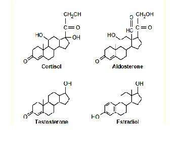
Structural Formula of Steroid Hormone
2. Steroid Hormone: This type of hormone is made up of steroid substances. It is usually a hormone secreted by the adrenal cortex (cortisol, aldosterone), ovaries (estrogen and progesterone), testis (testosterone) and placenta (estrogen and progesterone).
3. Amino acid derivatives or biogenic amines: These hormones are derived from amino acids such as melanin and adrenal medullary adrenaline.
4. Iodinated amino acids: Iodine is combined with amino acids to produce iodinated amino acids such as thyroxine.
The nervous system and the endocrine system existing in the animal body coordinate the functions of different systems. So these two systems are called integrated systems. Hormones are important regulators that often regulate the metabolism of different types of living cells. The endocrine gland is unanimously referred to as the ductless gland whose secretory substance is released directly into the blood or lymph. Based on the origin, the components of the endocrine system are divided into the following:
(1) Discrete endocrine glands: Such glands are pituitary (hypophysis), thyroid and pineal.
(2) Organs containing exocrine and endocrine functions: In fish, kidneys, gonads and intestines perform such functions. Heterotropic thyroid follicles, interenal and stannius corpuscles are present in the kidneys.
(3) Scattered cells with endocrine function: These are called expanded neuroendocrine. These cells are found in the digestive tract. These are usually called paracrine cells (such as somatostatin). These are gastrointestinal peptides that are classified as specific hormones or paracrine agents. They are also called Pupative Hormones.
The organs that contain endocrine glands are called endocrine organs. They are present in the internal organs. Almost all types of endocrine glands are present in the fish like in the upper vertebrates but their tissue and physiology are not clearly known. Although the endocrine glands have similarities with the upper vertebrae, some differences can be observed due to living in the aquatic environment.
Although mammalian endocrinology is more advanced, but studies have been performed on hormone-regulating functions in fish. These studies have shown that pigment cells, the effect of hormones on germ cells, the function of the pituitary and thyroid glands, balance in movement, nitrogen metabolism (adrenal cortical tissue, thyroid gland), maturation of sex cells and reproductive behavior(pituitary galnd and sex hormones), the effect of hormones on metabolism (adrenal cortical tissue) have been proven.
In elasmobranchii and osteichthyes, the complexity of the endocrine glands have been shown to increase. The elasmobranch has well-developed endocrine glands but differs slightly compared to the higher chordetss. The endocrine glands of the osteichthyes are more similar to the higher chordates. The endocrine glands in fish and mammals differ due to the development and metaformosis of different systems.
The endocrine glands of mammals are more advanced and well-known, but the endocrinology of fish is limited to the effects of hormones on chromatophores, the function of sex cells, the activity of the pituitary and thyroid glands, and the control of osmosis. The function of the endocrine system is not like that of the nervous system.
The endocrine system mainly controls the relatively slow metabolism of carbohydrates and water through the adrenal cortical tissue, the metabolism of nitrogen through the adrenal cortical tissue and thyroid gland, and the maturation and sexual behavior of germ cells through the pituitary and sex hormones.
There are 12 types of endocrine glands in fish. Their names are mentioned below:
- Pituitary gland
- Thyroid gland
- Interrenal tissue
- Chromaffin tissue
- Corpuscles of stannius
- Ultimobranchial gland
- Islets of Langerhans
- Gonads
- Intestinal mucosa
- Thymus gland
- Pineal body
- Urophysis
The pituitary gland is the most significant among the endocrine glands. This gland secretes hormones directly on their own and regulates the function of other glands by helping them secrete hormones. Hormones cannot work alone. Hormones have to rely on certain external stimuli such as light, temperature and water chemistry to function. All these activities are performed under the direction of the brain.
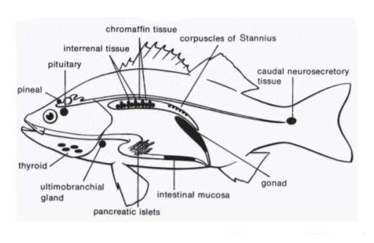
Image showing location of endocrine glands in fishes
Pituitary gland or Hypophysis
The pituitary gland is called the master gland. This is because the hormones secreted by these glands directly through the help of different biochemical reactions and reveals various characteristics, as well as play a role in controlling the secretion of other endocrine glands. This gland is found in all types of vertebrates. The anterior end of the pituitary gland is adjacent to the optic kyazma and the posterior end is located in the saccus vasculosus. This gland is attached to the roof of the Dianecephalon by the infandibulum.
The pituitary gland is mainly divided into two parts based on the cell structure, relative position, presence of secretory cells and pigmentation capacity of different parts of the pituitary gland, viz.
(1) Adenohypophysis
(2) Neurohypophysis
Adenohypophysis): It is divided into three types: These are:
- Pro-adenohypophysis
- Meso-adenohypophysis
- Meta-adenohypophysis
Pro-adenohypophysist: It covers most of the frontal lobes of adenohypophysis. It is also called rostral pars distalis. It contains acidophilic cells which are stained with azocarmine and orange G. In some fish species, a significant number of such acidophilic cells, such as follicular structures, surround the blood capilaries. In addition to acidophils, there are some cyanophils that are stained with aniline blue or aldehyde fuchsin (AF).
Meso-adenohypophysis: It exists between pro-adenohypophysis and meta-adenohypophysis. It is also called proximal pars distalis. Meso-adenohypophysis cannot be precisely distinguished from pro-adenohypophysis. However, as a result of the proliferation of multiple branches of the parenchyma (Neurohypophysis), it can be easily distinguished from meta-adenohypophysis. Meso-adenohypophysis decreases in size at different times of the year. It contains acidophils, cyanophils and chromophobes. Cyanophils is seen as the main ingredient during fish maturation and spawning. The number of acidophils is high during the rest of the year. Acidophils is pigmented with azocarmine or orange G pigment. However, cyanophils are aniline blue, Periodic acid-Schiff (PAS), aldehyde fuchsin (AF) and aldehyde thionin. Two types of cyanophils are found in most fish species based on size, shape and coloring ability. Chromophobes do not accept any pigments.
Meta-adenohypophysis: It is usually located in the posterior part of the hypophysis. It is also called Pars Intermedia. It partially or completely covers the distal edge of the neurohypophysis and connects to the meta-adenohypophysis, forming numerous branches from the neurohypophysis. Its size varies widely among different species. Its cells can be divided into acidophils and cyanophils based on their pigmentation capacity. However, some species also contain cells called amphiphils, which are stained with aniline blue and orange G. Moreover, there are some chromophobes here. Cyanophils in this region exhibit negative reactions to Periodic acid-Schiff (PAS), aldehyde fuchsin (AF) and aldehyde thionin.
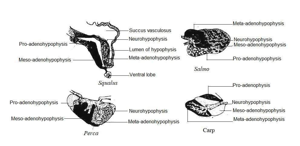
Image showing pituitary glands of different fishes
Neurohypophysis
Numerous nerve fibers originating from the cells of the hypothalamus enter the pituitary gland and re-branch into meta-adenohypophysis. These two tissues are so closely related that they are considered together as neuro intermediate lobes. The nerve fiber of neurohypophysis in the Elasmobranch contains pituitary and neurosecretory elements or particles which are stained by aldehyde fuchsin (AF), chrome alum, hematoxylin and aldehyde thionin. Some teleosts have lymphocytes and migratory cyanophils in this region. Large grains of neurosecretory material are sometimes called herring bodies. This area of the hypophysis is glandular. Due to the presence of two types of cells, it is thought that they are involved in the production of two types of hormones.
Histophysiology of the Pituitary Gland and Various Hormones
Hypophysectomy and mammalian hormone injections show that the pituitary gland in fish produces nine different types of hormones like mammals. All hormones are made up of proteins or polypeptides. The hormones are-
- Prolactin Growth hormone (somatotropin; STH)
- Gonadotropins (GTH) – These are two types: Follicle stimulating hormone (FSH) and luteinizing hormone (LH)
- Thyroid stimulating hormone (TSH)
- Adrenocorticotropin (ACTH)
- Melanocyte stimulating hormone (MSH) or intermedine
- Vasopressin and
- Oxytocin.
Recent studies have shown that potential areas and cell types are responsible for the release of these hormones. The following is a description of these cells:
Prolactin cells: These cells are mainly located in pro-adenohypophysis and meso-adenohypophysis or pars distalis and form dense mass. In some species, such cells have a follicular structure. Such cells contain red pigments in erythrocytes or azocarmine and the acid fuchsin. However, they are not pigmented with Periodic acid-Schiff (PAS), Alkan Blue or Aldehyde fuchsin (AF). Studies have shown that these cells secrete prolactin-like hormones.
Function: Prolactin affects milk production in mammals, but in fish it plays a role in controlling circulation.
Corticotropes (ACTH cells): Such cells are also located in pro-adenohypophysis and neurohypophysis (NH). They are thought to secrete adrenocorticotropic hormones. In fish such as Poecilia, these cells are granular long chromophobic types. Sometimes these cells are lightly pigmented with erythrocyte or azocarmine and turn gray with lead hematoxylin. In eel fish, these cells contain more grains which are more pigmented in alizarin and lead hematoxylin.
Function: The hormone produced from these cells stimulates the adrenal cortical tissue to produce ACH and metabolize stored fats, water, carbohydrates and proteins. These cells also contain sodium ions and help in the metabolism of electrolytes. In Carassius fish, it helps in cell melanogenesis.
Somatropes (STH cells): It is formed from acidophils cells of pro-adenohypophysis and meso-adenohypophysis and is pigmented by orange G. Some species of teleost have two types of acidophil cells. One type of cell is pigmented by orange G (somatotropes). Other types of cells are stained with azacarmine or erythrosine (prolactin cells). In some species, such as Anguilla and Poecilia, two types of acidophils cells have been distinguished based on location, morphology, and pigmentation capacity. In many teleosts, such as Anguilla and Poecilia, the somatotropes are acidophils type and are located in the meso-adenohypophysis and are mixed with thyrotropes.
Function: Such cells help to increase body size.
Gonadotropes (FSH and LH cells): These cells are usually present in meso-adenohypophysis, but sometimes, especially during the sexual maturity of eel (Olivereau 1967) and trout (Baker 1968), these cells enter pro-adenohypophysis. These cells contain glycoproteins and are stained by PAS, aldehyde fuchsin (FA), alcian blue and aniline blue. Various studies have shown that two types of gonadotropes are found in fish which are known as FSH and LH in mammals. Specific physiological and biochemical tests have shown that these two types of hormones are secreted by teleosts but not readily available (Olivereau 1967). The relationship of gonadotropes with sexual perfection has been proven experimentally on Indian teleost.
Function: These cells control the secretion of sex hormones.
Thyrotropes (TSH cells): Thyrotropes and gonadotropes are types of basophils (mucoid cells). They are found in meso-adenohypophysis. Thyrotropes cannot be distinguished from gonadotropes in most teleosts. However, there are differences in stained capacity. In some teleosts such as Phoxinus, Rutilus and Astyanax, thyrotrophs aldehydes are used to stain fuchsin (AF). Gonadotropes, however, are not stained by AF. Moreover, their positions vary. Thyroid hormone (TSH) is present on the surface of meso-adenohypophysis and in some species of such as Poecilia, gonadotropes are found in the ventral side of meso-adenohypophysis.
Function: Controls the secretion of thyroid gland.
The presence of two distinct cells in meta-adenohypophysis suggests that the region secretes two types of hormones. One of these is the melaminophore stimulating hormone (Intermedin-MSH) and the other is the melanophore concentrating hormone (MCH-Melanophore Concentrating Hormone).
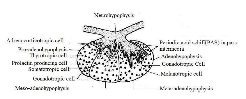
Image Showing of Hormone Secretory Cells of Pituitary glands in Fishes
Neurohypophysis
It releases two types of hormones, such as arginine vasotocin and oxytocin.
Functions: Arginine plays an important role in controlling vasotocin secretion and maintaining salt-water balance, and oxytocin helps in sexual intercourse and egg laying.
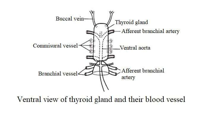
Thyroid Gland
In most teleosts, the thyroid gland is composed of numerous follicles surrounding the arterial artery and the afferent bronchial blood vessel. However, in some fish such as Parrotfish (Spariidae), Swordfish (Xiphias gladius), Gymnarchus niloticus and Thynnus thynnus it is densely packed and covered. Some aerial fish such as Channa punctatus, C. gachua, C. marulius, C. striatus and Heteroneustes fossilis have a dense and covered thyroid gland near the aortic artery (Belsare 1959). Intermediate conditions are observed in Clarias batrachus. In this case, the gland does not have a capsule, the gland seems to be three condensed grains.
Histologically, the thyroid gland is made up of numerous follicles. The follicle, like a hollow ball full of fluid, contains a single layer of epithelial cells. These follicles vary in size and shape and are joined by connective tissue. As this gland is more rich in red blood cells, the blood supply system is well developed in it. The epithelium around the follicle may be thin or thick, and the height of the cells depends on their excretory activity. Normally less active follicles contain thinner epithelium. There are two main types of cells in the epithelium. These cells are-
(1) Chief cells – These cells are dense and columnar in shape and have transparent cytoplasm attached.
(2) Colloid cells (Colloid cells or Benstay`s cells) – These cells contain granules of excretory material.
In some teleosts, the presence of thyroid follicles in various abnormal locations such as the main kidney, brain, esophagus and pleura has been reported. The idea is that all these heterotropic thyroid follicles migrate from the pharyngeal region to different parts of the body. The thyroid follicle collects inorganic iodine from the blood and stores it in its cells. It then combines iodine with tyrosine to make thyroid hormone. These hormones are secreted from the thyroid gland under the influence of thyrotropic hormones secreted from the pituitary gland. There are three types of thyroid hormones in fish, viz
- Mono-iodothyrosine
- Diiodotyrosine and
- Thyroxine.
Functions
(1) Thyroid hormone plays an important role in oxygen uptake. However, the current study has found the opposite result. In Goldfish (Carassius auratus), Muller (1953) has been shown to increase their oxygen uptake by applying the hormone thyroxine, but Matty (1954) has shown that removal of the thyroid gland in the elasmobranch (surgical thyroidectomy) has no effect on respiration. In the case of teleost, the application of thiouria has been shown to reduce the need for oxygen uptake.
(2) Thyroid glands affect the control of osmosis (salmon and Gasterosteus).
(3) It plays a role in the growth of fish (goldfish) and nitrogen depletion.
(4) This gland, in combination with other endocrine glands, influences the migration of fish.
(5) Hormones in the thyroid gland play a role in the metabolism of carbohydrates in fish. Studies have shown that when the thyroid gland is activated, the concentration of glycogen in the liver decreases. Applying thiourea reduces liver glycogen. According to some researchers, the thyroid activity increases during the sexual maturity of fish.
Interrenal Tissue
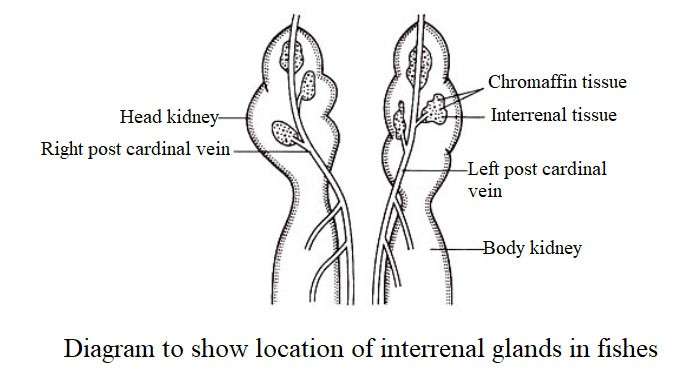
It is also called adrenal cortical tissue. In Lamprey (cyclostomata), endocrine internal cells are present throughout the body cavity adjacent to the posterior cardinal vein. In the case of Rays, it is located near the posterior renal collar. In the case of sharks (Squaliformes) it is located in the middle of the kidney. In most teleost fish, internal tissue is found in the main kidney near the posterior cardinal vein. Internal cells are located in one or more rows along the veins. In some fish, such as Puntius ticto, the interenal cells form a thick granular lump. In Channa punctatus, on the other hand, the cells form small lobules and are arranged along the posterior cardinal vein. These cells (internal cells) are eosinophilic in nature. Different species of teleost vary in the structure and extent of internal tissue. These tissue secrete two types of hormones, viz
- Mineral Corticoid Hormone – It plays a role in regulating circulation.
- Gluco-corticoid hormone – It affects the level of blood sugar.
Steroid-converting enzymes, primitive steroids, and corticosteroid hormones have recently been found in the interenal tissues of fish. Hypophysectomy results in atrophy of the intracranial cells of some fish. Studies have shown that the interenal tissue of the teleost is hmologous part of the mammalian adrenal cortex and produces cortical steroids. The secretion of interenal tissue is regulated by adrenocorticotropic hormones of the pituitary gland.
Cromaffin Tissue
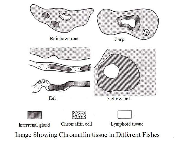
This is called suprarenal bodies or medullary tissue. In the lamprey (Cyclostomata) chromafin cells are located along the portal veins of the dorsal aorta, ventricle and heart in the form of a thread. In sharks and rays (Elasmobranchii) these nerves are located in the sympathetic chain of the nerve ganglia. It differs greatly in the extent of chromafin tissue (adrenaline-producing) in teleost. This tissue is adjacent to the intra neural collar. In some teleosts, chromafin cells are scattered in the main kidney. These form singles or groups within the adrenal cells or form adrenal complexes jointly with the adrenal glands. The chromafin tissue of the fish is given below:
- Adrenaline;
- Norepinephrine;
- Di-hydroxyphenylalanine (`dopa`) and
- 5-Hydroxytryptamine (serotonin).
Functions
(1) When chromafin tissue extract is injected into other fish, it stimulates the sympathetic nervous system.
(2) The hormone produced from chromaffin tissue concentrates the pigment granule in melanophore.
(3) It controls blood pressure.
(4) It acts in the same way as the mammalian adrenal medulla, so the chlomafin tissue of the teleost is considered to be a homologous organ of the adrenal medulla of mammals.
Corpuscles of Stannius
In 1839, a scientist named Stannius first described the presence of this particle in the kidneys of sturgeon as a distinct glandular structure. These are small nodule-shaped structures that are partially or completely hidden on the dorsal, lateral, or anterior side of the bony fish. They are oval or round in shape and the number of cells in different species varies from 1-6. It is usually located in the posterior part of the kidney and is thought to be an endocrine organ. From an embryological point of view it originates from the pronephric or mesonepric tube (Belsare 1973).
In some fish, especially goldfish, trout, salmon, etc., this particle is found to be flat, white, and oval in shape on the peritoneal surface. From histological point of view, each stannius particle consists of parenchymal cells. They are separated by connective tissue and surrounded by fibrous capsules. There are also vascular ganglionic units that contain ganglionic cells, blood vessels, and the nervous system, and are located near or inside the parenchyma cells (Belsare 1973).
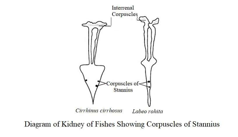
AF + and AF– are found in salmon, Astyanax mexicanus and Anguilla anguilla (Nadkarni and Gorbman 1966, Rasquin 1965, Hanke and Chester Jony 1966). According to Belsare (1983), some teleosts such as Labeo rohita, Cirrhina chirrhosus (= Cirrhina mrigala), Catla catla, Clarias batrachus, Channa punctatus, etc., contain only one type of cell in stennius corpuscles. According to Gill and Punetha (1977), the mountain river Pseudoecheneis salcatus has 1-2 stannius corpuscles located 10 mm away from the excretory genitalia. The stannius are covered by the connective tissue capsule and contain a type of AF + secretory cell along the blood vessel.
Depending on the change in the salinity of the water, the grouping of cells decreases or increases. Hypophysectomy also results in loss of granules. Removing stannius particles from Anguilla anguilla reduces the amount of plasma sodium and increases the concentration of potassium and calcium. Moreover, it can kill the fish.
Studies have shown that the release of stannius particles affects the kidneys, alters kidney function, and maintains electrolyte homeostasis. Some researchers have suggested that the stennius particle is a coexistence of the adrenal glands of the spinal cord and they want to establish it as a posterior adrenal gland.
Table: Number, size and position of stannius in some older teleosts (After Belsare 1973)
|
Fish Species |
Number of Stannius Corpuscles |
Size (mm) |
Location* |
|---|---|---|---|
|
Labeo rohita |
2-3 |
3-6 |
50-60 mm |
|
Catla catla |
2 |
2-5.4 |
30-40 mm |
|
Cirrhina mrigala |
2-3 |
1-3 |
40-50 mm |
|
Clarias batrachus |
2-10 |
0.5-1.2 |
4-30 mm |
|
Heteropneustes fossilis |
2-4 |
0.4-0.9 |
20-25 mm |
|
Mystus vittatus |
2-4 |
0.15-0.25 |
10-15 mm |
|
Channa punctatus |
1-2 |
0.2-0.8 |
10-20 mm |
|
Notopterus notopterus |
1 |
0.2-0.6 |
Anterior part of the kidney |
* Distance in millimeters from the excretory genital pore
Ultimobranchial Glands
This gland is small in size and paired and is located on the horizontal membrane or near the thyroid gland in the space between the abdominal cavity and the sinus venosus on the left side of the esophagus. Trout fish exist as a band of white collars on the membrane. From an embryonic point of view, this gland develops from the pharyngeal epithelium near the fifth gill arch. It is histologically similar to the parathyroid glands of the upper vertebrae. In 130-150 mm length of heteropneustes fossilis, this gland is 0.471.5 mm (Belsare 1974).
From a histological point of view it is composed of parenchyma tissue. It is made up of solid and polygonal cell clusters covered by cell cords and capillaries. These glands secrete calcium-regulating hormone calcitosin (thyrocalcitosin). This hormone controls hypercalcemia and hypocalcemia. It is thought that this hormone also plays a role in regulating secretion. High concentrations of calcitonin are found in these glands of fish, but their specific function in the lower vertebrae is not known. The function of this gland is regulated by the pituitary gland.
Islets of Langerhans
The endocrine glands of the pancreas condense into numerous islets. These islets may have distinct entities and may extend to the pancreatic lobe. The large islets that are visible to the naked eye are called the main islets. Fish have more than one islets. They are usually located near the gallbladder, spleen or pyloric system. The islets in Channa punctatus are located near the spleen and pylorus. It is the largest of all islets. Moreover, some small islets are scattered in the mesentery. Each islet is covered by a thin layer of connective tissue.
The islet of spleen is usually covered by epidermal cells. The connective tissue trabeculae of all islets contain numerous small blood vessels. In Labeo rohita, Cirrhina mrigala, Schizothorax plagiostomus, the main islet is located near the bottom of the alimentary canal where the bile duct is exposed. Some other large and small islets are located in the mesenteric arteries near the gallbladder and bile ducts. The largest islet in the Tor tor is pin in shape, located near the bile duct. It is surrounded by extracellular glands and connective tissue. Some islets of carp are also covered by extracellular cells.
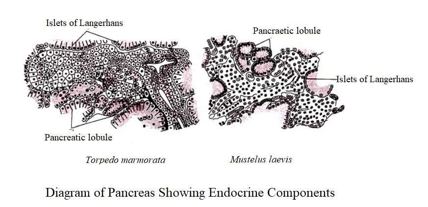
In Clarias batrachus, Heteropneustes fossilis, and Mystus seenghala, the exocrine glands are more precise, and the largest part is located in the mesentery along the blood vessels or bile ducts between the two lobes of the liver. The extracellular mass contains a significant number of pancreatic islets of various shapes. Islets are found near the gallbladder and bile ducts. All the islets are covered by the epidermis and are seen in the pancreas as small swellings. The Wallago attu contains specific pancreas that is located within the liver. The small islet is scattered among the mass of the cell, separated by a thick connective tissue.
In Mastacembelus armatus, it has a white macroscopic main islet in the duodenal loop near the front of the spleen which is covered by a thick connective collar capsule and it separates itself from the epidermal cells.
Histology
The islet glands of fish are relatively large in size and consist of three types of cells. Beta (B) cells in the cells are involved in the secretion of insulin and in aldehyde fuschin (AF) pigment, it shows a dark purple. AF-cells are called alpha (A) cells. These are of two types, namely: A1 and A2. Of these, A1 cells are silver positive i.e. it shows black color in silver pigment while A2 cells are silver negative. Fish have more A1 cells than A2 cells. Some researchers have referred to the agranuloid cells of some fish as C cells and A2 cells as D cells. Beta cells are usually located in the center of the group of islets, but some fish have two types of cell scattered throughout the islet. Beta cells are polyhedral-shaped cells with large spherical nuclei. Their cytoplasm is AF+. Alpha cells are triangular in shape with abnormal nuclei and their cytoplasm is AF–.
Beta cells secrete insulin in fish. However, as a result of hyperglycemia, beta cells are destroyed by taking beta-cytotoxic drugs such as aloxacin and streptozotocin. Glucose injections in fish increase blood sugar levels. In Heteropneustes fossilis, Channa punctatus and Clarias batrachus, spontaneous degranulation and degeneration of beta cells occurs. One of the two alpha cells secretes the hormone A1 glucagon. Some researchers believe that A2 cells secrete gastrin-like material or a lipocyclic-like substance. In fish (Heteropneustes, Clarias, Channa) hyperglycemia occurs when glucose is applied and hypoglycemia occurs when insulin is applied, resulting in degradation of beta cells.
Gonads
Fish have a pair of testes and ovaries known as gonads. The sexual maturation of fish and the development of gametes take place in the presence of sex hormones. Moreover, secondary sexual characteristics such as coloration, reproductive tubercles, maturation of gonopodia, etc. depend on the hormones secreted by the sex glands. The sperm, which resembles an open folded sac, produces spermatocytes. It is covered by the germinal layer. In addition to the main sex organ, the testes of fish act as endocrine glands.
The testes are made up of numerous seminiferous tubules and stromal or interstitial tissues. Ledig cells or interstitial cells secrete testosterone, a hormone called androgen, from the interstitial space, the secretion of which is regulated by GTH. Androgens play a stimulating role in the development, maturation and function control of the male sexual organs and in the process of spermatogenesis. Androgens work on the central nervous system and affect male sexual behavior. These hormones help in the metabolism of proteins and carbohydrates.
The female fish has a pair of ovaries in its abdomen which is known as the main female sexual organ. The ovaries contain numerous ovarian follicles and stromal tissues. The ovaries produce two types of steroid hormones, estrogen and progesterone. Estrogen stimulates the growth of secondary sex organs in female fish and promotes the growth of developing ovarian follicles. Moreover, estrogen controls the sexual behavior of the female fish.
Intestinal Mucosa
It regulates the production of pancreatic juice by secreting a hormone called secretin (pancreatozymen). This hormone is synthesized from the anterior region of the small intestine. Secretin affects the enzyme-carrying juices from the pancreas and accelerates the flow of zymogens. Secretin was first discovered by Bayliss and Starling (1902). They prepare secretin extract from the intestines of salmon, dogfish and skates.
Thymus Gland
The thymus gland is located on the dorsal or ventral side of the gill arch. It is a long and lobed organ. This gland is formed from the dorsal angle of the gallbladder as a solid external growth. It is rich in blood vessels. As the connective tissue enters the gland, it forms acinus. It is thought that the hormone thymosin secreted by this gland plays a role in the growth of the body.
Pineal organ
It is located near the pituitary gland. Researchers have studied the structure and function of this organ (Omara and Oguri 1969); Haferz 1972; Srivastava and Dubu 1975; Sastry and Sathynesen 1981, Khanna et al. 1983). The posterior-mid-dorsal growth of the epithelium from the posterior of the habenular produces this organ that passes through the anterior surface through the adipose connective tissue. It contains pineal sac, pineal thalamus and pineal stalk.
In some fish, pineal sacs touch the roof of the cranium. This is not the case with others. This attachment is thick at Atherenopsis and thin at Mystus aor. The pineal sac forms a capsule covered by collagen connective tissue. In some species, the pouch is hollow but in lantern it is solid. In different fish, the pineal sacs vary in size and shape. In Mystus aor, it resembles sagitate, in Bagarius bagarius it resembles a rosette or rose, in Wallago attu it is angular and in Nangra punctata it is funnel-shaped. The pineal sac is a light-sensitive organ in fish, but it also acts as an endocrine gland. Pinealectomy in Lebistes showed a decrease in their growth rate and abnormalities in the stimulation of the skeletal system, thyroid, pituitary and stennius corpuscles.
The pineal gland secretes melatonin hormone and sulfate acid mycopolysaccharides, basic proteins (proteins arginine and lysine amino acids), common lipids, acidic lipids, phospholipids, basophil-rich neurosecretory components containing gamma components. Moreover, evidence of the presence of metallic elements and components such as chromium, nickel, chromium phosphide, copper, aluminum copper, etc. in the pineal organ has been found (Srivastava et al. 1984). Recent studies on Mystus vittatus shown that the pineal organ affects the maturation of fish gonads. Some researchers have identified the pineal gland of fish as a light-sensitive and secretory organ.
Urophysis
Urophysis is a small oval structure located on the marginal part of the spinal cord. It is a storage organ that releases substances produced in the neurosecretory cells of the spinal cord. These cells of urophysis are called caudal neurosecretory system. Such mechanisms are found only in the elasmobranch and teleost, but they are similar to the hypothalamus neurosecretory system in vertebrates. In the caudal neurosecretory system, neurosecretory cells extend to the margins of the spinal cord. Neurosecretory cells are large nerve cells that contain basophilic cytoplasm and polymorphic nuclei. In carp and neoplasms, urophysis has a distinct oval structure.
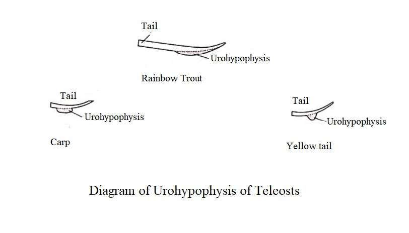
Urophysis is composed of components of the spinal cord such as neurosecretory axons, glia, glial fibers and meningeal derivatives such as tubular reticulum and reticular fibers. The idea is that the caudal neurosecretory system is involved in the control of osmosis. This gland secretes two peptide hormones, urotensin-1 and urotensin-2. Hormones thus play a role in regulating circulation, gas metabolism, buoyancy, corticotropin release, and smooth muscle contraction. The secretion from urophys is able to compress the smooth muscles of the ovaries and fallopian tubes of the guppy fish (Poecilia reticulata) and the sperm of the Gallichthys mirabilis. It is thought that it also plays a role in reproduction and spawning.
Table: Names of different types of endocrine glands of fish, their location, origin, secreted hormones, function and chemical nature
|
Name of Gland |
Location |
Embryonic origin |
Hormone |
Functions |
Chemical nature |
|---|---|---|---|---|---|
|
1. Pituitary |
|
|
|
|
|
|
(1)Posterior lobe-Neurohypophysis |
Base of the brain |
Ecto and mesoderm |
Arginine vesotocin |
Controlling osmosis and maintaining salt-water balance. |
Peptide |
|
Oxytocin |
Helping to have sex and lay eggs. |
Peptide | |||
|
(2) Middle lobe or region-meta-adenohypophysis |
Intermedin (MSH) |
Assistance in melanogenesis (Fundulus) |
Peptide | ||
|
Melanophore Concentrating Hormone(MCH) |
Melanin concentrates to reveal body color. |
Peptide | |||
|
(3) Transition lobe or meso-adenohypophysis |
Growth hormone inhibitory hormone(GHIH) |
Inhibiting the growth of the body. |
Peptide | ||
|
Growth hormone releasing hormone (GHRH) |
Assists in the growth of the body. |
Peptide | |||
|
Thyrotropin(TH) |
Control of thyroid gland secretion. |
Peptide | |||
|
Gonadotropin(LH,FSH) |
Controlling the secretion of sex hormones. |
Peptide | |||
|
Prolactin |
Assistance in melanogenesis with MSH. |
Peptide | |||
|
(4) Anterior lobe or region-Pro-adenohypophysis |
Prolactin |
Assists in electrolyte control and melanogenesis. |
Peptide | ||
|
Corticotropic releasing hormone(CRH) |
stimulates the pituitary synthesis of ACTH |
Peptide | |||
|
Adrenocorticotropic hormone(ACTH) |
Assistance in adrenal cortical secretion control and melanogenesis. |
Peptide | |||
|
2. Thyroid gland |
In bony fish it is located along the arterial artery, in cartilage fish along the midline of the tongue muscle |
Mesoderm and endoderm |
Thyroxine |
Role in accelerating sexual maturation, metabolism control, osmosis control and metamorphosis. |
Amine |
|
3.Ultimobranchial glands |
It is located esophageal ventral wall between esophagus and sinus venous |
Endoderm |
Calcitonin |
It plays a role in calcium metabolism. |
Peptide |
|
4. Suprarenal (chromafin tissue) |
Located on the dorsal side along the kidney |
Mesoderm |
Epinephrine |
Concentration of pigment grains in melanophore. |
Amine |
|
Norepinephrine |
Controlling blood pressure and pupillary swelling. |
Amine | |||
|
5. Adrenal cortical tissue or internal tissue |
Along the hemopoietic tissue-cardinal vein of the main kidney |
Mesoderm |
Cortisol |
Use of stored fats, protein formation and carbohydrates metabolism. |
Steroid |
|
Adrenal cortical steroid (aldosterone) |
Acceleration of water metabolism, sodium ion retention, electrolyte metabolism. |
Steroid | |||
|
6.Pancreatic islets |
It is located at the inner wall of larvae of lamprey, the hepatopancreas; In most bony fish, it is scattered in the pancreas. |
Mesoderm |
Insulin |
Metabolism of carbohydrates |
Peptide |
|
Glucagon |
Metabolism of carbohydrates |
Peptide | |||
|
7. Gonads |
Body cavity |
Mesoderm |
Testosterone |
Develops the reproductive system. |
Steroid |
|
Androgen |
Development of secondary sexual characteristics in men; |
Steroid | |||
|
Estrogen |
Accelerating the growth and development of the female reproductive system; Development |
Steroid | |||
|
Progesterone |
Little is known about the functions in fish. Stimulates pancreatic secretion. Electrolyte maintains |
Steroid | |||
|
8. Intestinal mucosa |
Small intestine |
Endoderm |
Secrtine |
Stimulates pancreatic secretion. |
Peptide |
|
9. Corpuscles of Stannius |
Posterior part of the kidney |
Mesoderm |
Unknown |
Maintaining electrolyte homeostasis. |
Unknown |
|
10. Thymus |
Dorsal edge of the gill arch |
– |
Thymosin |
Assist in physical growth. |
– |
|
11.Pineal body |
Diencepahlon |
– |
Melatonin |
Influence on melanophore. |
– |
|
12.Urophysis |
It is located at the end of the spinal cord |
– |
Eurotensin-1 and Eurotensin -2 |
Control of water and ion metabolism. |
Peptide |
Table: Difference Between Endocrine and Exocrine Glands
|
Endocrine Gland |
Exocrine Gland |
|---|---|
|
Since the endocrine gland does not have a duct, it is called ductless gland. |
Since the duct of the exocrine gland exists, it is called duct gland. |
|
Its secretion is called hormones (such as somatotrophin, testosterone, thyroxin, etc.). |
Its secretion is called non-hormonal chemicals (such as sweat, milk, sebum, enzymes, etc.). |
|
Its secretion flows directly through the blood. |
Its secretion flows through the duct. |
|
These glands are located inside the body. |
These glands are located inside and outside the body. |
|
It operates in the target region, far from the place of origin. |
It operates near the point of origin or in the distant target area. |
|
In animals, such glands are pituitary, thyroid, parathyroid, gonad, etc. |
Such glands in animals are sweat glands, breast glands, salivary glands, sebaceous glands, stomach, liver and pancreas. |
Table: Difference Between Hormone and Enzyme
|
Hormone |
Enzyme |
|---|---|
|
Hormones are secreted from the endocrine glands. |
Enzymes are secreted from the exocrine glands. |
|
Hormones are called chemical messengers. |
Enzymes are called organic catalysts. |
|
Hormones reach the destination either directly or through the blood. |
The enzyme is transported from the place of origin through the duct to the destination. |
|
Hormones are made up of peptides and proteins or amines or steroids or fatty acids derivatives. |
The enzyme is made up of globular proteins. |
|
Hormones are destroyed after biochemical reactions. |
Enzymes are not destroyed through biochemical reactions. |
|
A hormone is capable of influencing one or more reactions. |
An enzyme is only able to act as a catalyst in a particular reaction. |
|
They are dynamic and arise in one place and act in another. |
They are located in all the cells and act in that place. |
|
It plays a role in controlling physical growth or decline, color, and sexual characteristics etc. |
It controls all the biochemical reactions in the cell. |
