
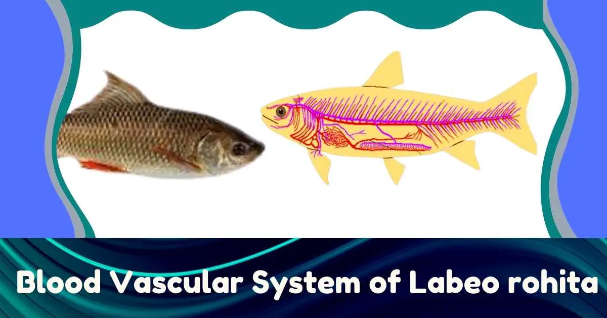
The blood circulatory system of fish is of closed type. Blood circulates in the body from being confined to specific ducts. Like other bony fish, the carbon dioxide-rich blood is collected through veins from the various body parts of the Rohu fish (Labeo) and first accumulates in the heart.
This blood then enters the gills to enrich the oxygen and from there the blood does not return to the heart but circulates directly to different parts of the body. That is, the blood collected from the body tissue enters the heart and from there it spreads to different parts of the body in a single flow path. Since such a flow completes a cycle, hence, it is called a single circuit circulation.
On the other hand, in all cases from amphibians to mammals, carbon dioxide-rich blood enters the respiratory tract (lungs) from the body to the heart to enrich oxygen and then returns to the heart and from there spreads to different parts of the body. In this case, the blood circulates through the heart in two separate cycles, hence, it is called double circuit circulation. The circulatory system of Rohu fish consists of blood, the heart, arteries, veins, and capillaries.

Blood
The blood of Rohu fish is slightly paler in color than that of terrestrial vertebrates. Three types of blood corpuscles are floating in colorless plasma. Red blood corpuscles (RBC) are ovoid, bifurcated, nucleated, and rich in hemoglobin. White blood cells (WBC) have no definite shape and no nucleus. The platelets are nucleated and spindle-shaped.
Heart
The heart of Rohu fish is located in the pericardial cavity at the base of the body. The heart lies under the pharynx behind the gill pouch. The heart is covered with a pericardium membrane. Their heart is divided into three chambers: A sinus venosus, an auricle, and a ventricle. The heart of Rohu does not have Conus arteriosus. However, the ventral aorta swells to form the bulbus arteriosus.
Sinus Venosus
Sinus venosus is located on the surface of the heart. It is a thin-walled sac-like chamber. Carbon dioxide-rich blood collected from different parts of the body enters the sinus venosus through the anterior cardinal vein, posterior cardinal vein, inferior jugular vein, hepatic vein, and subclavian vein.
All collected blood enters the auricle from the sinus venosus through the sinu-auricular aperture. There is a pair of valves in the sinu-auricular aperture.

Auricle
The auricle is located in front of the sinus venosus and above the ventricle. Although the auricle is oval, its outer part is lobulated. Its wall is thin and the muscles are underdeveloped. The auricle is exposed to the ventricle with the auriculo-ventricular aperture. There is a pair of valves in this aperture.
Ventricle
This is the largest chamber of the heart. It is located below the auricle. It looks almost like pear-shaped. Its wall is quite thick and highly muscular. The inner wall contains a muscular ridge called columnae carneae and an elastic cord called Chordae tendineae. This is why the inner part of the ventricle shows unevenness. The ventricular cavity is exposed in an opening through the balbus arteriosus. A pair of crescent-shaped valves is placed in this opening.
Bulbus Arteriosus
It is a flask-like muscleless sac that is located in front of the ventricle. It is not the chamber of the heart. Because it does not have cardiac muscle. The bulbous arteriosus is associated with the ventral aorta.
Circulation of Blood Through Heart
The blood enters the gills from the heart to be oxygenated. This is why the chambers of the heart contract at regular intervals. At first, shrinking occurs in the sinus venosus. Then the auricle and finally the ventricles contract. Thus blood flows to the sinus venosus > anus > ventricle > balbus arteriosus > the ventral aorta. However, the valve located at the entrance of each chamber obstructs the reverse flow of blood. As a result, the blood flowing occurs from the back only to the front, never reversing occurs. The heart of a fish is called the branchial heart or venous heart.

Carbon dioxide-rich blood is collected from different parts of the body and stored in the sinus venosus. The sinus venosus contracts when it is full of blood and the blood enters the auricle through the sinu-auricle aperture. At this time, when the auricle contracts, blood enters the auricle through the auriculo-ventricle aperture. At this time the sinus venosus expand but the blood can never go back in the opposite direction. As the ventricular muscle contracts, blood enters the balbus arteriosus and from there the blood enters the gills through the ventral aorta through the afferent branchial artery.
Arterial system
The blood vessels that usually carry blood to a place far from the heart are called arteries. And all the arteries together form the arterial system. However, the arteries of fish are a little different. Some of these arteries carry blood from the heart to the gills, and some arteries collect blood from the gills and transmit it to different parts of the body. The arteries that carry blood from the heart to the gills are called afferent branchial arteries.
On the other hand, the arteries that collect blood from the gills are called efferent branchial arteries. Then the arterial system of Rohu fish consists of afferent and efferent branchial artery, dorsal aorta and its branches.
Afferent Branchial Artery
The ventral aorta originates from the heart’s balbus arteriosus and moves forward through the middle of the gills on both sides along the floor of the pharynx. From the ventral aorta, four pairs of afferent branchial arteries are generated and enter the four pairs of gills on either side.
The anterior part of the ventral aorta splits to produce the first pair afferent branchial artery and a little behind the second pair the afferent branchial artery. They enter the first and second pairs of gills, respectively. The third and fourth pairs of afferent branchial arteries located behind the second pair of arteries originate from both sides of the ventral aorta and enter the third and fourth gills, respectively. They divide into capillaries after supplying blood to each gill lamella.

Efferent Branchial Artery
A special feature of the circulatory system of fish is that oxygen-rich blood from the gills does not return to the heart but is transmitted directly to different parts of the body through the efferent branchial arteries. Oxygenated blood from the gills travels through four pairs of efferent branchial arteries to different parts of the body. The first and second efferent branchial arteries connect laterally to each other to form a lateral aorta.
The two lateral aortae on either side combine to form the dorsal aorta. A cephalic artery forms at the base of the first efferent branchial artery and divides into internal and external carotid arteries. The internal carotid supplies blood to the brain and the external carotid supplies blood to the eyes, tongue, and hyoid. The lateral aortae join the carotid artery in the anterior pharyngeal region to form an oval ring. Its name is Circulus cephalicus. The dorsal aorta extends posteriorly along the ventral midline of the vertebral column and forms the following branches.

Subclavian artery: The dorsal aorta originates from two sides of the subclavian artery and supplies blood to the thoracic girdle and dorsal fins.
Coeliaco-mesenteric artery: Originating from the aorta, this artery supplies blood to the swimbladder, the alimentary canal, the liver, and the spleen.
Renal artery: A few pairs of renal arteries protrude from both sides of the posterior part of the dorsal aorta and enter the kidney.
Iliac artery: The dorsal aorta protrudes a few pairs of iliac arteries from the very back and delivers blood to the pelvic fins.
Caudal artery: The dorsal aorta narrows the posterior region and supplies blood to the caudal region in the form of caudal arteries.
Venous System
Carbon dioxide-rich blood from different parts of the body returns to the heart through veins. Blood is collected from the anterior cardinal and inferior jugular veins from the front of the body of Rohu fish and enters the ductus cuvieri. At the same time, carbon dioxide-rich blood reaches the ductus cuvieri from the posterior part of the body through the posterior cardinal vein. Ductus cuvieri are exposed in sinus-venosus.
Carbon dioxide-rich blood from the posterior region of the body and the digestive tract is carried by the hepatic portal vein to the liver. This vein terminates in the capillary blood vessels in the liver. Blood then accumulates in the sinus venosus from the capillary blood vessels through the hepatic veins.
The caudal vein collects carbon dioxide-containing blood from the caudal region and divides it into two branches after entering the body. A branch bends to the right and moves forward as a right posterior cardinal vein. The other branch bends to the left and divides into many branches in the kidney. This is called the renal portal vein.

The left posterior cardinal vein originates from the kidney. This vein moves forward. Some segmental veins collect blood from different parts of the body and supply it to the right and left cardinal veins. The posterior cardinal veins connect horizontally through the transverse vein in some of them to form a transverse anastomosis. Subclavian veins collect blood from the thoracic fins and bring it to the sinus venosus. Note that Rohu fish(Labeo rohita) do not have an abdominal vein.