The heart is a special pump device with a valve in the circulatory system. In case of fish, the heart is a folded tube that contains three or four enlarged areas. The blood brought through the veins travels from the heart to the gills through the ventral aorta and the heart always contains carbon dioxide (CO2) or unrefined blood. This is why the heart of a fish is called the venous or branchial heart.
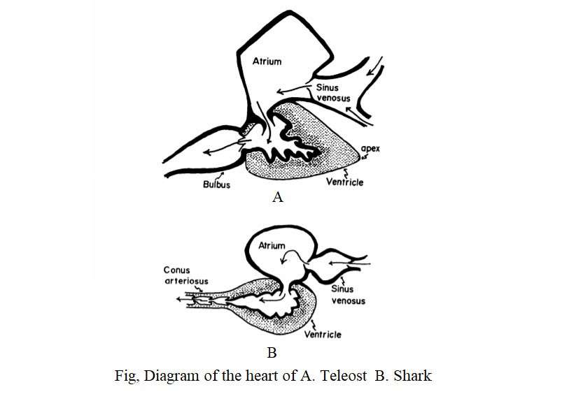
Blood passes through the aortic arch on the front and enters the gills to exchange gaseous components through the heart. In most fish, the heart is located immediately after the gills. In the case of the teleost, the heart is located at the front side of the body rather than at Elasmobranch. The heart is primitive type in Elasmobranch among the fish. It is located in the pericardial cavity and consists of the sinus venosus, atrium, ventricle and well-developed contractile conus.
Some researchers consider the atrium and ventricles to be the chambers of the heart. Some researchers also consider the sinus venosus and conus arteriosus to be the chambers of the heart. In the case of fish, there is some controversy over Conus arteriosus and Bulbus aorta. The fourth chamber of the elasmobranch is known as the conus arteriosus. In Teleost, however, it is known as Bulbus arteriosus.
The difference between conus arteriosus and bulbus arteriosus is that conus arteriosus has ventricular like heart-muscle and innumerable valves are continuously arranged in it, but bulbus arteriosus consists only of smooth muscle fibers and elastic tissue. (Boas 1980; Smith 1918; Danforth 1912; Parson 1929; Karandikar and Thakur 1954).
According to Torrey (1971), a teleostian fish (Cyprinus carpio) has both conus and bulbus arteriosus. According to Kumar (1974) and Santer (1977), the teleost has only bulbus arteriosus. On the other hand, Elasmobranch and Agnatha have conus arteriosus instead of bulbus arteriosus.
The heart has sino-auricular and sino-ventricular openings that are controlled by a dual valves. The conus has six rows of valves. Muscular and contractile conus are considered to be of primitive nature. It is found in some lower teleosts such as Acipenser, Polypterus and Lepidosteus.
In addition to conus, bulbus arteriosus exists in the Amia. It originates from the fibrous wall of an uncontrollable region. This type of secondary condition is seen in some lower teleost (Clupeiformes). Albula, Tarpon and Megalops have distinct conus and rows of transverse valves. The size and weight of the heart varies with the body weight of the fish. The structure of the heart of different fish is described below:
Heart of Cyclostomes
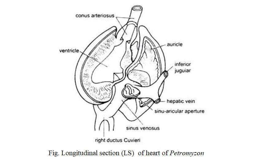
Lamprey’s (Petromyzon) heart is like the English letter ‘S’. It is formed by folding the posterior side of the gill and the sub-intestinal vessels. The larval heart develops as a straight duct. Later, this duct becomes longer and takes the shape of ‘S’ in a limited space. The heart consists of the sinus venosus, an atrium and a ventricle, and the conus arteriosus and is covered by the pericardium. A cartilage plate holds the pericardium. The sinus venosus is a thin-walled chamber that is exposed through a sinus-auricular opening to a thin-walled atrium at the top. The atrium is again connected to the thick-walled ventricle through the auricle-ventricular opening.
Heart of Cartilaginous (shark) Fishes
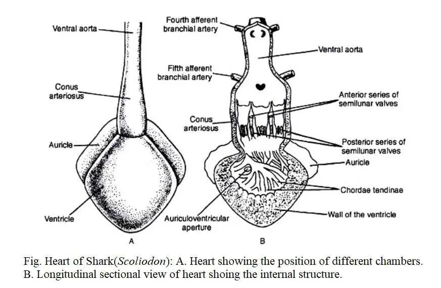
Their heart is a curved muscular duct that consists of the receiving region and the transmitting region. The receiving region consists of a sinus venosus and dorsally located an atrium while anterior portion contains a ventricle and a conus arteriosus. The heart is covered by a membrane called the pericardium. The dorsal part of the pericardium is made up of basibranchial cartilage. The heart is located between the two rows of gill pouches on the ventral side of the body of the fish.
Heart of Bony Fishes
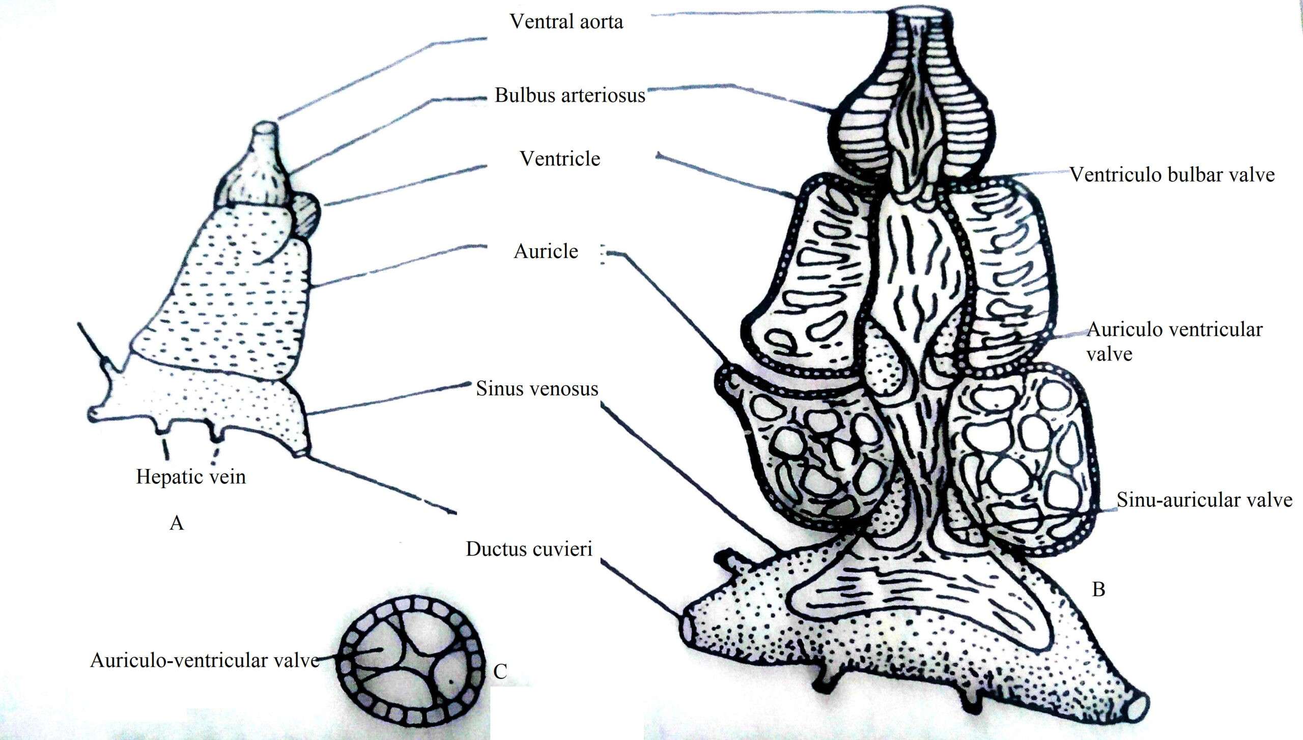
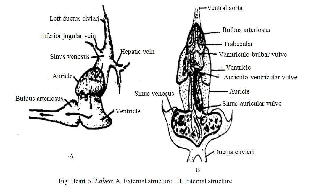
(a) Heart of Tor tor : The heart is located at the tip of the septum transversum in the pericardium sac. It consists of sinus venosus, atrium, ventricle and bulbus arteriosus. The sinus venus is a smooth-walled chamber that receives blood supply through the joint Ductus cuvieri, the joint hepatic vein, a posterior cardinal, and an inferior jugular vein. The openings of these blood vessels have no valves. The sinus venosus is exposed to the atrium through the sinus-auricular opening. In this opening, a pair of membranous semilunar valves are present. Each valve has a long wing pointed towards the front of the atrium.
The atrium encloses the ventricle dorsally and is relatively large in size with an irregular outer surface. It is orange and sponge-like soft and has a narrow cavity extending to the ventricles. The spongy wall of the atrium has numerous spaces or cavities covered by muscle fibers extending in different directions. The atrium-ventricular opening contains two pairs of semilunar valves of approximately equal shape. Each valve has a short wing adjacent to the atrium wall and a long wing adjacent to the ventricular wall, but the extended part of the valves points towards the atrium.
Fig. A. Heart of Tor tor, B. Internal structure of heart of Tor tor C. TS of auricular-ventricular aperture showing two pairs of valve
The ventricle is a superior muscular chamber with a thick wall and a narrow cavity. It is associated with bulbus arteriosus through ventricular-bulbar opening. There are a pair of semilunar valves in this opening. Each valve has a short wing adjacent to the ventricular wall and a long wing adjacent to the wall of the bulb so that the wings cross each other. The valves are hanging in the ventricular cavity. The wall of the bulbus is thin and has a narrow hole in it. In its cavity, a thin ribbon-like innumerable trabeculae pass parallelly. The bulbus extend into the ventral aorta anteriorly.
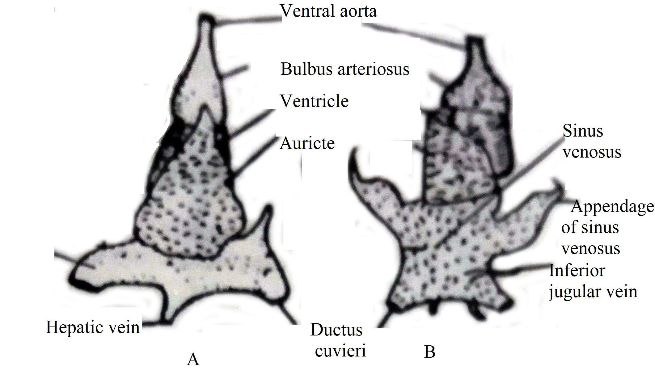
(b) Heart of Other Teleost: The heart of cyprinids such as Labeo rohita, Cirrhina mrigala, Catla catla and Schizothorax has the same general structure as Tor tor. Lebeo rohita, Cirrhina mrigala, and Catla catla have large sinuses and a pair of lateral appendages (Singh 1960). In the first two species, it is spongy and fibrous.
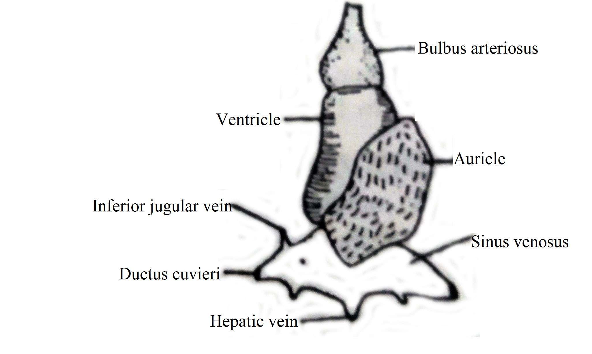
In Clarias batrachus, Mystus aor, Wallago attu, the sinus venosus is a thin-walled chamber in which a pair of membranous sinus-auricular valves are located obliquely along the long axis of the heart. One end of the dorsal valve extends to the front and reaches the atrium cavity and is attached to it.
Fig. Showing Heart of A. Wallago attu, B. Catla catla
Fig. Showing Heart of Clarias batrachus
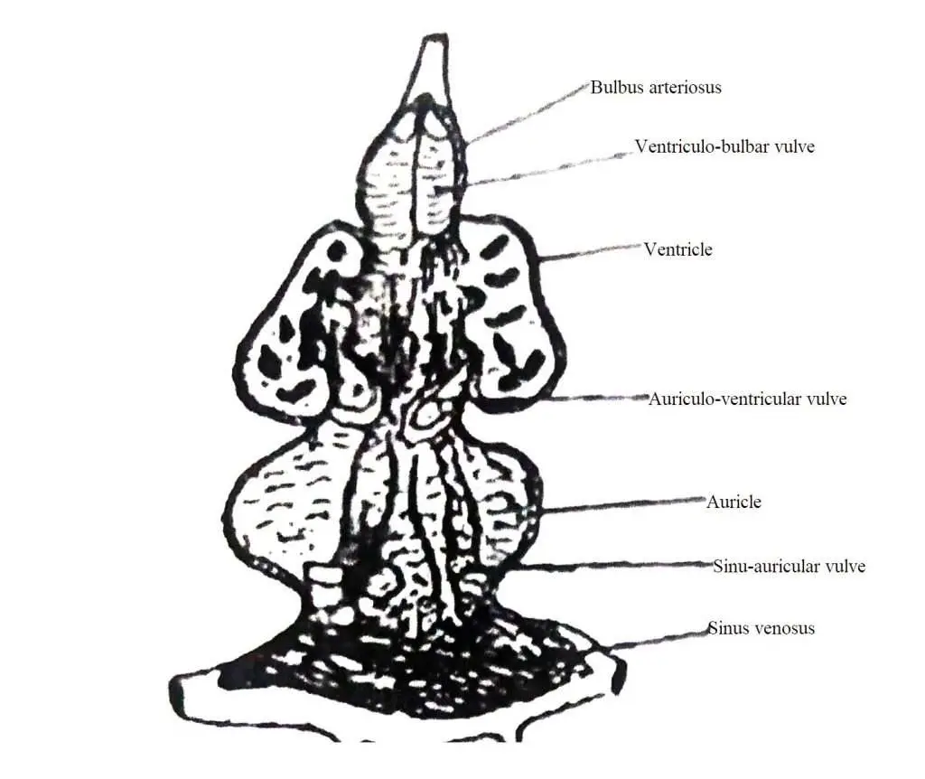
The atrium is structurally spongy type, looking like a beehive. The opening of the atrium ventricle has four valves, two of which are well-developed, while the other two are small, not so important. The ventricle has advanced muscular and two ventriculo-bulbar valves which are semilunar-like in shape. In case of Channa striatus, sinus venosus is small and there is no sinus-auricular valve.
Notopterus notopterus has 5-7 nodular valves in the sinus-auricular opening and Chitala chitala has 8-10 valves. Two of the 4 auricular-ventricular valves are small in size. Chitala chitala has a muscular conus arteriosus between the ventricles and the bulbus. In case of Notopterus, the ventricular bulbar valve is like a ribbon with a strange structure and divides the bulbous cavity into three chambers by a pair of vertical septum.
Working of the Heart
The venous blood travels to the heart, reaches the sinuses applying pressure to the semilunar valve, and reaches the atrium. During this time, the pockets of the valves are filled with blood and the pressure created by the contraction of the atria causes the valves to swell and obstruct the flow of blood from each other.
Due to the pressure of the four auricular-ventricular valves, the blood reaches the ventricle from the atrium and as soon as possible the ventricular cavity is filled with blood. During this time, the valves receive blood. So the valves swell and close the openings from being firmly attached to each other. As a result, the reverse flow of blood is obstructed. The blood then enters the bulbus by applying pressure to the ventriculo-bulbar valve. Inside the bulbus, the blood pressure rises again, causing the valves to swell and close the passageway, obstructing the retrograde flow of blood, causing the blood to flow forward through the ventral aorta.
Cardio-vuscular Control
Fish control the cardio-vascular system in two ways, viz
(1) Aneural and
(2) Neural mechanisms.
Aneural cardio-vascular control is accomplished through the direct response of the heart muscle to changes in temperature and the secretion of various glands and changes in blood volume. Temperature acts as an anural regulator due to the direct action of the myocardium on the pacemaker. In some species, an increase in temperature increases the heart rate, resulting in higher cardiac energy. By increasing blood flow, it is able to supply more oxygen to the body. As a result, higher metabolic rate is possible in warm water. Anural control also occurs under the influence of certain hormones such as epinephrine stimulates heart rate.
Neural control techniques occur through the tenth carotid nerve (vegus). The heart of these fish is nerved by a branch of the vegus nerve. Stimulation of the vegus nerve reduces the heart rate in the elasmobranch and teleosts. Different types of stimuli such as flashing light, sudden movement of an object, touch or mechanical vibration reduce the heart rate in fishes. In responding to environmental or other changes, fish face some problems during maintaining their blood circulation balance.

