
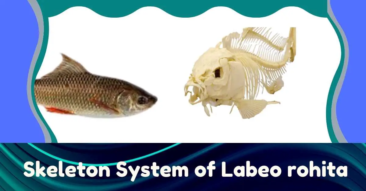
Labeo rohita (Rohu Fish) is a Actinopterygian fish (=Osteichthyes) which belongs to the family Cyprinidae under order Cypriniformes. It is a popular sports and food fish due to its high commercial value. The epidermis is located on the outside of the skin of the Rohu fish and the dermis is located on the inside. The epidermis contains pigment cells and mucus glands. Mucilage is secreted from the mucous glands, which keep the body moist, disinfected, aids in respiration, and facilitates movement. Scales are produced from the dermis. Their scales are cycloid in nature. Rows of scales are vertical, but arranged angularly. The back of a scale is covered by the front of the next scale. The transparent part around the center of the scales is called the focus. Some circular lines can be seen around the focus. Their name is circulus. Through these lines, knowledge about annual growth can be gained. The muscles are located under the skin. The myotome is zigzagged in the body and tail muscles.
Endoskeleton of the Labeo rohita
The endoskeleton of the Rohu fish is made of bones. Their endoskeleton is mainly divided into axial skeleton and appendicular skeleton.
Axial Skeleton
The axial skeleton consists of the skull and vertebral column.
Skull
The skull of a Rohu fish consists of a cranium, Sensory capsule, and visceral arch. The sensory capsule is inextricably linked to the cranium. However, the bond between the cranium and the visceral arch is not strong.
Cranium: The cranium of the Rohu fish is a bit long. Of these, the orbito-temporal, occipital, and otic regions are the most notable. The roof of its orbito-temporal region consists of two pairs of dermal bones called frontal and parietal. The frontal part contains the frontal, orbito-sphenoid, and parasphenoid bones.
")
The parasphenoid forms the floor of the cranium. This bone is quite long. It extends from the back of the vomer to the pro-otic bone. The parietal part contains the parietal and elisphenoid bones. The posterior part of the cranium is called the occipital region. There are three types of bones in this region: namely supraoccipital, basioccipital and exoccipital. There are three openings in the back of the occipital region. One is the middle foramen magnum and the other two are ovoid fenestra. This fenestra expands by perforating the occipital bone.
")
This is one of the unique features of the cranium of Cyprinid fish. The occipital spine is located on the surface of the supra-occipital. The occipital bone contains a spherical part called the occipital condyle, which connects the skull to the vertebral column.
")
Sensory Capsule: The otic region consists of a pair of auditory capsules. This capsule covers the inner ear. There are four bones in the otic region of Rohu fish. These bones are Pre-otic, Epiotic, Sphenotic, and Terotic Bone (Opisthotic bone is present in other teleosts). These four bones together form an inverted cup-like structure.
The nasal region of the skull contains the nasal and nostril bones. The bones of this region are nasal, ectoethmoid, lacrimal, mesethmoid, vomer, and rostral.
Visceral Arch: The seven pairs of semicircular bones are placed on either side of the pharyngeal wall and they are connected to each other along the midline and form seven visceral arches.
The first visceral arch is called the mandibular arch, the second is called the hyoid arch and the remaining five are called a branchial arch. The first four branchial arches contain four gills. And the fifth branchial arch forms the inferior pharyngeal bone. This bone is transformed into a masticating plate, which consists of large rods.
The first visceral arch or mandibular arch forms two jaws. The upper jaw contains three cartilage bones called palatine, metapterygoid and quadrate and two dermal bones called ectopterygoid and endopterygoid. The two premaxillae meet at the very front of the mouth. And there are two maxillae on either side of its back part. Both the premaxilla and the maxilla are dermal bones.
The upper jaw is connected to the cranium partly through the palatine and partly through the hyomandibular and symplectic. Each half of the lower jaw is composed of the small articular, large dentary, and small angular bones. The two dentaries are connected to each other along the midline.
The second visceral arch or Hyoid arch is divided into two parts. Hyomandibular is placed at the top and Hyoid cornu at the bottom and they together form the hyomandibular suspensorium, through which the jaws hang along with the cranium.
The posterior five visceral arches are called branchial arches. The four branchial arches on the front contain four gills. The fifth branchial arch is a reduced type. Each branchial arch is transformed into four replacement bones on each side. These are Pharyngobranchial on the surface, Epibranchial on the side, Ceratobranchial, and Hypobranchial on the ventral side.
The hypobranchial on both sides is connected to a medial basibranchial at the base. All the basibranchial and basihial come together to form a median ventral bar on the floor of the pharynx.
The lid-like operculum is present in the gill chamber of Rohu fish. The operculum consists of a combination of four bones. The largest bone in the operculum is called the Opercle. It looks triangular, wide, and flat. Its surface is convex with growth lines. There is a focus on its ventral side. It is attached to the skull by a condyle which is located at the tip.
The crescent-like and flattened bone is present on the front of the opercle which is known as the preopercle. It covers some parts on the front of the opercle. The third bone below the posterior edge of the opercle is called the subopercle.
It looks like a flattened sword. Its front part is narrow and the back edge is wide. The fourth bone is called the inter-opercle. It is located between the pre-opercle and the sub-opercle bones. It has a large groove on the underside.

Vertebral Column
The vertebral column of Rohu fish consists of 37-38 vertebrae. The vertebrae are bony and amphicoelous in nature (concave at both ends of the centrum). The vertebral column is divided into two regions. There are 21 vertebrae in the front part of the trunk region. And there are 16-17 caudal vertebrae in the posterior part of the tail region. The structure of the vertebrae in the trunk is different from that of the tail or caudal vertebrae.
Trunk Vertebrae: In the embryonic state, the vertebrae of the trunk have a narrow duct in the center of the double-concave centrum through which the notochord extends. In the adult state, the duct closes. From the front side of the centrum, two neural prophyses are formed on both sides, forming a neural arch around the spinal cord.

From the surface of the neural arch, a long neural spine rises upwards and extends backward. From the base of the neural arch to the anterior, a pair of small and blunt prezygapophysis originates and another pair of postzygapophysis originates posteriorly. Two short transverse processes or parapophyses extend from the ventral side of the centrum.
The pleural or dental ribs are connected to the parapophyses by ligaments. Ribs surround the abdominal cavity from the sides and back.
The first four vertebrae of the trunk are transformed into small bones or ossicles. None of them have parapophyses, but they have two transverse processes of each. From the first vertebral neural arch, two small bones originate, called Claustrum and Scaphium. They are the most frontal part of the Weberian ossicles. The second and third vertebral centrum are joined together to form a flattened triangular bone on each side, the outer side of which is like a slice of the moon. It is called Tripus.
It is the very back of the Weberian ossicle. There is a small bone called the intercalarium on each side between the scapula and the tripus. It represents the neural arch of the second vertebra.
The claustrum, scaphium, intercalarium, and trypus join together by ligaments to form a chain of ossicles. The scaphium edge of this chain connects to the inner ear and the tripus to the swimbladder, helping the sound waves felt in the swimbladder or air sac to reach the inner ear.

Caudal Vertebrae: There are 16-18 caudal vertebrae in the caudal region of the vertebral column. Their structure is roughly similar to that of the trunk vertebrae. However, two parapophysis-like basapophysis emerge from both sides of the centrum and merge along the ventral midline to form the Haemal Arch.
The Haemal spine emerges from the Haemal arch and bends backward. The surface of the caudal vertebrae has a neural arch and neural spine and the ventral part has a haemal arch and haemal spine. Two posteroventral processes are formed on both sides from the base of the Haemal arch. The posterior blood vessels dilate through the Haemal Arch. There are no ribs attached to it.

The last three caudal vertebrae change to contain the caudal fin and at the same time provide firmness. A partially bony notochord is attached to the last caudal vertebrae to form a rod, which extends upwards. This is called urostyle.
The haemal arches of the last three caudal vertebrae form broad hypurals, and their neural arches combine with wide epurals to form a vertical plate-like structure so that the caudal fin rays or Lepidotrichia are bound together and at this time it looks like fans.

Appendicular Skeleton
The skeleton of pectoral fins and pelvic fins and the corresponding pectoral and pelvic girdles form the appendages of the fish.
Pectoral Girdle
The pectoral girdle is located behind the last branchial arch. The two bony cycles on either side are separated from each other. Each pectoral girdle consists of three cartilage bones (e.g., scapula, coracoid, and mesocoracoid) and four dermal bones (cleithrum or clavicle, supracleithrum, post-temporal, and post-cleithrum.

Scapula: The scapula is a spherical bone, located on top of the coracoid, which is also an irregular triangular bone. This bone lies in the ventral side of the mesocorcoid in the scapular bone. Mesocoracoid is inverted ‘Y’ shaped. The glenoid facet is located between the scapula and the coracoid. Through this, three of the four radials of the pectoral fins are connected performingly.
Pectoral Fin
Pectoral fins are supported by 19 fin rays or Lepidotrichia. Lepidotrichia is connected with four radials. And the scapula maintains a direct connection with the radials.
Pelvic Girdle
The pelvic girdle is made up of a single bone on each side in front of the anal fins. This bone is known as basipterygium. The front end is forked and the rear end is attached to pelvic cartilage.

The forked end is connected to the twelfth trunk vertebral rib by a ligament. The two pelvic girdles on either side join each other in the midventral surface.
Pelvic Fins
Each pelvic fins carry nine fin rays. The fin rays are attached to the three radials located behind the basipterygium. The first two radials carry two fin rays while the third radial contains the remaining fin rays.