
Pinus are coniferous, evergreen resinous trees which are popularly known as pine. They belong to the family Pinaceae under order Coniferales of class Coiniferosida. Different species of the genus Pinus are distributed throughout the temperate and sub-alpine regions of Northern Hemisphere where they form dense forests of evergreen trees. They can grow up to 80 (260 ft) meters in height. The tallest pine tree is ponderosa pine which can grow about 81.79 m (268.35 ft) tall. They are long lived plant and can live up to 1000 years or more under favorable conditions.
There are about 120 species of pine tree throughout the world. They are widely distributed in the hills and have economic value because many pines are used in the construction, paper-products industries as the source of timber and resin. Some pine trees are also sources of wood tars, resin, turpentine, and oils while some produce edible seeds such as pine nuts, piñons, or pinyons. Besides these, white, black, Himalayan, and stone pines are cultivated as ornamental pine trees.
Systematic Position
Division: Coniferophyta
Class: Coiniferosida
Order: Coniferales
Family: Pinaceae
Genus: Pinus
Species: P. wallichiana, P. insularis, P. armandi, Pinus khasya, Pinus geradiana, Pinus roxburghii, etc.
You may also read: Gymnosperms: Salient Characteristic Features
Salient Features of Pinus
- They are evergreen, perennial lofty trees with spirally growing branches which give pyramidal or conical appearance.
- The body is divided into stem, roots and needle-like leaves.
- The stem is erect and cylindrical and is cov¬ered with bark.
- There are two types of branches: the long shoot of unlimited growth and dwarf shoot of limited growth.
- The long shoot bears apical bud and grows indefinitely with many scaly leaves.
- Dwarf shoot does not contain any apical bud and they arise on the long shoot in the axil of scaly leaves.
- Each dwarf shoot bear two scaly leaves which is also known as prophylls.
- Leaves are dimorphic: the long green needle shaped foliage leaves and small, brown, membranous scale leaves.
- Scale leaves are thin and brownish in color which is developed only on long as well as dwarf shoots while the foliage leaves are large, needle-like and found only at the apex of the dwarf shoots.
- The pine bears tap root system with insufficient hairs but it disappears soon. Many lateral roots also develop which play an important role to absorb the mineral containing water.
- The branch roots are infested with mycorrhizal fungus and hence it is called the mycorrhizal root.
- They have endarch vascular bundles. Individual vascular bundles are separated by means of medullary rays.
- The anatomy of leaves shows xerophytic structure: thick cuticularised epidermis with sunken stomata and sclerenchymatous hypodermis.
- Resin ducts are present in the mesophyll tissue and the cells of the mesophyll have ridges on the walls which project inside the cell cavities.
- Microsporophylls are arranged spirally on the central axis and forms male cone.
- Megasporophyll of the female cone is composed of large ovuliferous scale and lower smaller bract scale, which are the free from each other.
- Each ovuliferous scale bears two anatropous ovules or megasporangia.
- The pollen grains are winged.
- During the development of male gametophyte, two prothalial cells are formed which later on degenerates. Besides these, 2-3 archaegonia are formed with a neck of eight cells.

External morphology of Pinus
The plant body represents the sporophyte. The sporophytes are evergreen and tall tree (10 -80 m in height). The body has three parts: root, stem and leaves. The plants bear well developed tap root system. The stem is stout, branched and pyramidal in shape with recemose branches.
Root: A strong tap root system is present in young plant which may persist or roots develop and become stronger adventitious roots with increasing age. The roots can grow on rocks or hard ground and spread over a large area. The lateral roots are well developed with insufficient root hairs. Often a branch roots are infested with mycorrhizal fungus and hence it is called the mycorrhizal root.
Stem: The stem is erect, stout, cylindrical and pyramidal shape with dimorphic branches. The branches are restricted in the apical region. The stem is covered with bark. There are two types of branches:
Healthy Origins Pycnogenol (Nature’s Super Antioxidant)
The long shoot of unlimited growth: The main branches or long shoots have an unlimited growth with scale leaves which are found below the dwarf shoots and the needle like foliage leaves are present exclusively at their terminal ends.
The dwarf shoot of limited growth: The dwarf shoots develop in the axils of scale leaves on the main branches, which are without apical buds. It is about 1 -2 cm long with one or two scale leaves. The dwarf shoot also contains foliage leaves. In this case, a dwarf shoot with its foliage leaves is known as spur.
Leaf: The pine tree bears two types of leaves:
The scale leaves: Both long and dwarf shoots bear scale-leaves and fall off as the branches attain maturity. These leaves are small, brownish in color and membranous with protective structures.
The foliage leaves: The dwarf shoots bear foliage leaves. The leaves are long, green, simple, needle-like with photosynthetic structures. They develop in clusters at the apex of the dwarf shoots and can form the spur. Their number varies from 1-5 in different species.

Internal Morphology
Stem: A young stem in cross section resembles a dicotyledonous stem in many respects. It is wavy in outline. The general arrangement of various tissues from the periphery to the center is as follows:
Epidermis: It is the outer surface layer of the stem. It consists of a single layer of tubular close compact parenchymatous cells with a thick cuticle.
Hypodermis: Below the epidermis, a hypodermis layer is preset. It consists of a few layers of lignified sclerenchymatous cells.
Cortex: It consists of several layers of parenchymatous cells with copious resin ducts. A single layer of resin secreting glandular epithelial cells surrounds the resin duct.
Endodermis: Outside of the pericycle, endodermis is present. It consists of a single layer of parenchymatous cells.
Pericycle: It consists of several layers of parenchymatous cells which are located outer to the ring of vascular bundles.
Vascular bundles: They are conjoint, collateral, open, arranged in ring and endarch. In this case, protoxylem is directed towards the pith. Between xylem and phloem, a narrow strip of cambium is present. The xylem is composed of tracheids with bordered pits and xylem rays, without vessels. The phloem is composed of sieve tubes and phloem parenchyma without companion cells.
Pith: It is composed of a mass of parenchymatous cells.
Pith rays: It is composed by a narrow strip of cells running from the pith outwards between the vascular bundles.
")
Leaf: A transverse section of foliage leaf shows the following structure under the microscope. It is semi-circular in outline (centric type of leaf).
(i) Epidermis: It consists of a single layer of thick walled cells with a heavy sheet of cuticle and sunken stomata.
(ii) Hypodermis: It consists of two or three layers of very thick walled sclerenchymatous cells with resin ducts, which are broken up by stomata. It is placed just underneath the epidermis. It strengthens the tissue of the leaf.
(iii) Mesophyll: It consists of a few layer of chloroplast containing parenchyma cells with peg-like projections. In this case, wall projecting occurs inside the cell cavity which is known as arm palisade.
(iv) Endodermis: It consists of a single layer of barrel shaped cells which is present outside of the pericycle.
(v) Vascular bundles: They are collateral, closed and two in number.
(vi) Pericycle: It consists of albuminous cells and tracheidal cells. In this case, albuminous cells lie close to the phloem while tracheidal cells lie adjacent to the xylem. Albuminous and tracheidal cells together form the transfusion tissue which helps to flow of nutrients. The xylem is surrounded by pericycle.
")
Root: The internal organization of tissues in the root of Pinus is almost similar to that in a dicotyledonous root. A cross section of young root shows an epiblema (piliferous layer) with root hairs, a multilayered cortex and a diarch to pentrarch vascular cylinder and Y-shaped xylem bundles. 2-3 layered pericycles surround the vascular bundles while the cortex consists of a few layers of thin walled parenchymatous cells. A single layered endodermis is present which is followed by a pericycle. The root is mycorrhizal because fungus grows on the surface of the root.
Reproductive Structures of Pinus
The Pinus is monoecious plant which shows the sporophytic generation. The microsporophyll (male) and megasporophyll (female) are formed on the same plant but these two types of sporophylls appear usually in separate cones or strobilli.
The male and female cones are known as staminate strobilus and carpellate strobilus, respectively. The Pinus does not show vegetative reproduction. The flowers are unisexual. They always occur on the shoots of the current and a little away from the apex.
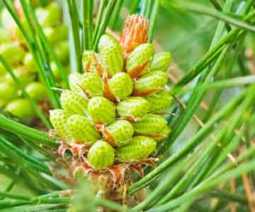
Male cone (staminate strobilus)
The male cones are simple, compact, oval structures and about 2-3 cm long. They are found to occur in clusters, near the tip of the long shoots.
Each male cone bears a short and elongated central axis upon which a large number of microsporophylls or stamens are arranged spirally. The microsporophylls are scaly and their number varies from 60 to 135 in each cone.
A microsporophyll consists of a short filament or stalk and a terminal leaf-like expanded structure while the apex is slightly bent upwards. Each microsporophyll bears two pouch-like microsporangia (anthers or pollen sacs) on its ventral surface. A microsporangium is sessile and oblong which is supported with a jacket of several layers of cells.
Each microsporangium produces a several microspores (pollen grains). The wall of each microspore is covered by inner intine and an outer exine. The microspores are winged and yellow in color. In this case, wings help in the dispersal of spores by wind.


Female Cone (ovulate strobilus)
The female cones are larger and compound in nature. They are formed in clusters of 1-4 in the axils of scale leaves of long shoots. Initially, they are green but ultimately become brownish red in color. It starts to produce in winter and become ready for pollination during the spring. It is hard woody and dry structure. It bears a central axis upon which a large number of megasporophylls are arranged spirally.
Each megasporophyll has short stalk with a large ovuliferous scale on the upper surface and a small bract scale on the lower surface. Each ovuliferous scale bears two inverted megasporangia on its upper surface towards the base.
Each megasporangium consists of a massive tissue which is called the nucellus and an envelope which is known as the integument. At the basal region, the integument is fused with the nucellus and open at the top by forming micropyle.
A single megaspore mother cell is differentiated within the nuceller tissue, which divides meiotically to form four megaspores. Of these four megaspores, only the lower most one is functional while others degenerate. The only functional megaspore increases in size and takes part in the development of the female gametophyte.

Female cones and foliage of Pinus contorta

Structure of the gametophytes
Male gametophyte: The microspore is the first cell of the male gametophyte. Microspore begins to germinate within the microsporangium and produces an extremely reduced male gametophyte. The microspore first divides to form a very small first prothellial cell and a large cell. The large cell then cuts off a second prothellial cell adjacent to the first one and the remaining of the large cell forms the antheridial cell. The two prothellial cells soon degenerate. The antherdial cell again divides to form a small generative cell above and a large tube cell below. The nucleus of tube cell is known as tube nucleus which regulates the growth of the pollen tube.
The microspores are dispersed with the help of wind. Further development of microspores takes place when it reaches the ovule due to pollination. A number of microspores reach the megasporangium (ovule) where they are attached with sticky mucillagenous substance from the micropyle.

Ovule of Pinus
When mucillagenous substance dries up, some of the microspores are drawn inside on to the apex of the nucellus. Later on, the spore coats split between the wings. Then the tube cell protrudes and grows to form the pollen tube. Ultimately, the pollen tube penetrates the nucellus. The generative cell then divides to form a sterile stalk cell and a fertile body cell (spermatogenous cell). The body cell further divides to form two non-motile unequal male gametes (sperms).
Female gametophyte: The functional megaspore is the first cell of the female gametophyte. It germinates within the megasporangium. The functional megaspore enlarges in size, its nucleus divides repeatedly by free nuclear divisions to form about 2,500 daughter nuclei. All the nuclei lie in the cytoplasm of the megaspore.
After that, a large central vacuole is produced, whereby the cytoplasm together with the nuclei moves towards the periphery. After pollination, development of the female gametophyte takes place again.
The walls are broken down around the nuclei and ultimately, a solid mass of thin walled cells are formed. The massive tissue thus formed within the megaspore. It is known as the female gematophyte. It is often referred to as the endosperm. The endosperm tissue is haploid (n).
A few flask shaped archegonia (2-3) are formed from the superficial cells (archegonial initials) that lie towards the micropylar end of the female gematophyte.
A mature archegonium consists of a neck of eight cells, one ventral canal cell and a large egg. There is no neck canal cell.
Fertilization
After a year of pollination, fertilization occurs. The pollen tube moves downwardly and reaches the neck of the archegonium. It then penetrates the neck and the tip of which bursts to discharge the two male gametes. One of the male gamete unites with the egg to form a diploid zygote.
New Sporophyte
The zygote is the first cell of the sporophyte which germinates almost immediately after its formation. The zygote nucleus divides to form four free nuclei. These nuclei then moves to the bottom of the zygote where they divide again to form eight nuclei. Subsequently, walls appear between these nuclei except the upper two. Further divisions result in the formation of sixteen cells in four tiers; this sixteen celled structure is termed as the proembryo.
The upper most tiers of four open cells merges into the general mass of the cytoplasm to carry out nutritive function. The next tier of four cells is called rosette tier which is capable of forming abortive embryo; normally it supplies nutrients to the suspensor and the embryo.
The third tier of four cells constitutes the suspension tier. The lower most tier of four cells is the embryo tier. The cells of the suspension tier elongate to form four separate suspensors, each carrying an embryo cell at its tip. The cells of the embryo tier form the embryos. The elongated suspensors push down the embryos into the gametophytic tissue and thus help the later to receive their nutrients.

Each cell of the embryo tier divides to form four potential embryos and secondary suspensor. Formation of more than one embryo in each megasporangium is referred to as polyembryony. In fact, only one of these embryos attains maturity and the others undergo degeneration.
After fertilization, the integument and the megasporangium are converted into seed coat and seed respectively. The seed contains embryo, the membranous nutritive layer perisperm and kernel. The nutritive tissue lies surrounding the embryo. A mature embryo consists of a radical, a hypocotyls, several cotyledons and a plumule. The seed germinates under favorable conditions to form a new sporophyte. This type of germination is called epigeal germination.
Final Words
Pine trees are widely distributed throughout the world and have lots of economic value. Pine tree wood is very strong and it is extensively used to manufacture doors, electric poles, window panes, boats, railway sleepers, musical instruments, boards, boxes, veneers and plywood, building construction, paneling, etc. due to its durability. Turpentine oil is produced from the pine tree which is used as a solvent for varnishes, paints and in perfumery industry. It is also used for producing disinfectants, synthetic pine oil, denaturants, and insecticides, etc. Pine oil from the pinewood is used in pharmaceutical industries, textile industries, leather industries, etc.
You might also read: Reproduction in Algae