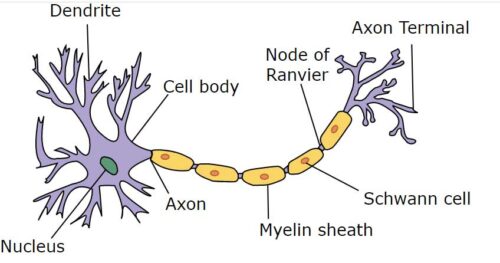
Nerves and specialized cells (neurons) comprise the nervous system. Structurally, the nervous system includes two parts such as CNS (the central nervous system) and the peripheral nervous system. The nervous system is highly specialized epithelium tissue for reception, discharge, and transmission of stimuli. The neuron or nerve cell is the structural and functional unit of the nervous system. . Specialized cells or neurons convey signals between different parts of the body. But the neurons of CNS are supported by several varieties non-excitable cells which together are called neuroglea.
Classification of Neuron
Neuron may be classified as follows:
According to function, neuron may be of three types:
- Afferent (sensory) neuron
- Efferent (motor) neuron and
- Intercalated neuron.
According to number of axon and dendrites from the cell body, the neuron are the following types:
(i) Apolar neuron: When there is no axon or dendrite are present but contain only cell body, it is called apolar neuron.
(ii) Unipolar neuron: When one axon and one dendrite combined and originates from one pole of the cell body, the neuron is known as unipolar neuron.
(iii) Bipolar neuron: In this type of neuron, one axon and one dendrite originates from opposite poles of the cell body.
(iv) Multipolar neuron: In this type, many dendrites and one axon arise from several parts (poles) of the cell body.
Structure of Neuron
A neuron consists of the following two parts:
- Cell body
- Processes
Cell body
The cell body often called cyton or centron or parykarion. A typical cell body may be rounded or polygonal or spindle shaped or flask-shaped or pyramidal shaped. The surface of the cell body is covered by fine plasma membrane. The cytoplasmic matrix of the nerve cell is known as neuroplasm. It contains a spherical nucleus with a large nucleoplasm.
Special bodies called Nissl granules, a network of the ultramicroscopic fibrils, the neurofibrils, Golgi apparatus, ribosomes, endoplasmic reticulum, centrosome, etc. are present in the neuroplasm. The cell body is present in the ganglia and central nervous system, i.e., in the gray matter of the brain and spinal cord. It regulates the functional activities of the neuron.
Processes
The cell body of the neuron gives rise several branching process which are of the following two main types:
- Axon and
- Dendrite
The anatomically the neuron contains the following parts:
- Cell body
- Dendrites
- Axon
- Nodes of Ranvier
- Axon terminal bundle
- Myelin sheath cells
Axon
There is usually a long branch of neuron, known as axon. It arises from a special protuberance on the cell body, known as the axon hillock. The axon is generally called the nerve fiber. The cytoplasm of the axon is known as axoplasma which is covered by a fine plasma membrane as axolemma. The axon is branched at it distal end known as axon terminals which make contact with other neuron to form synapse or with muscle to form neuromuscular junction.

Structure of Neuron: Image credit-wikipedia.org
Dendrites
Beside the axon there is one or shorter branched processes, called dendrites. The dendrites serve to carry message to the nerve cell. The sensory neuron has single, long dendrites instead of many dendrites. Motor neurons have multiple thick dendrites. The neuroplasm of the cell body also has a extension to each dendrite. All deddrites have synaptic knobs at the ends which are the connections to the adjoining nerves. The dendrite`s function is to carry a nerve impulse into the cell body. In the nervous system the cell body of the neuron combine to form the gray matter and all axons and dendrites combine to form white matters.
Myelinated and Non-myelinated nerve fiber
The axon, both in the vertebrates and invertebrates are often covered with a sheath called the myelin sheath such fibers are called myelinated fiber while those which are not covered are called non-myelinated fiber. Dendrites are always non-myelinated.
Myelinated Nerve Fiber
It is composed of three elements: axis cylinder, myelin sheath and neurolemma. The axis cylinder(axon) is the innermost part covered by the thin membrane axolemma and filled up with axoplasm. Surronding the axon is myelin sheath that is approximately as thick as the axon itself.
The myelin sheath provides both mechanical support and electrical insulation to the axon. The myelin sheath is broken at regular intervals along the length of the axon. The place where the myelin sheath is discontinuous are called Nodes of Ranvier. All nerve fibers outside the CNS (central nervous system) receive another homogeneous nucleated continuous covering the neurolemma or sheath of Schwann.
Functions of Neurolemma
- The neurolemma protects the nerve fiber
- It does supply nutrition and
- It helps in the repairing of damaged peripheral nerves.
Non-myelinated fiber
This type of nerve fiber does not contain myelin sheath but only axon cylinder and neurolemma are present. Hence the diameter of the nerve is less than myelinated fiber.
A single nerve is composed of bundle of several axons. The individual axon is covered by neurolemma. The connective tissue layer surrounding the individual,axon is called endoneurium. Several axons are bounded together by another connective tissue sheath called perineurium. Several sheath bundles of axons are combined together by another connective tissue layer called epineurium to form a nerve.
Neuroglea
It is spherical type of nerve tissue present in between the neurons. Neuroglea are three types such as:
- Astrocytes
- Oligodendrocytes and
- Microglia.
Functions of Neuroglea
- Support
- Insulation
- Pahocytosis
Difference between axon and dendrites
|
Axon |
Dendrite |
|---|---|
|
It is usually long and single process of neuron. |
It is usually short and multiple process of neuron. |
|
It has uniform thickness and unbranched. |
It is branched and tapper at the end. |
|
It is myelinated. |
It is non-myelinated. |
|
Characteristic 4 |
Characteristic 4 |
|
Neurolemma is present. |
Neurolemma is absent. |
|
Myelinated sheath is differentiated into nodes and internodes. |
Node and internodes are not differentiated. |
|
Structurally differentiated from the cell. |
Structurally same as cell. |
|
The external appearance is smooth. |
The external appearance is rough. |
|
It conducts impulses away from the cell body. |
It conducts impulse to the cell body. |
|
Each cell contains one axon. |
Each cell contains many dendrites. |
|
It does not contain robosomes. |
It contains ribosomes. |
