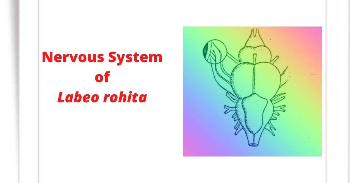
The nervous system of Rohu fish (Labeo rohita) is divided into the following three parts:
A. Central nervous system
B. Peripheral nervous system and
C. Autonomic nervous system.
A. Central Nervous System
The central nervous system is made up of the brain and spinal cord. The brain of the Rohu fish is divided into three regions:
- Forebrain (Prosencephalon)
- Midbrain (Mesencephalon) and
- Hindbrain (Rhombencephalon).
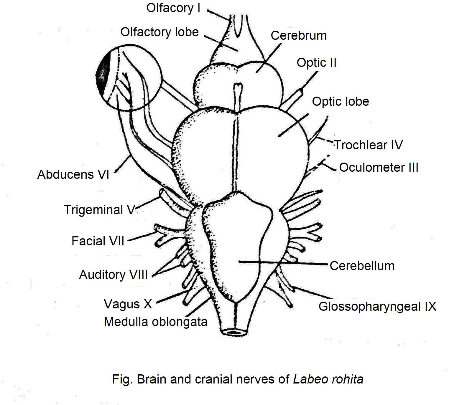
The forebrain is further divided into telencephalon and diencephalon. There are two olfactory lobes at the tip of the telencephalon. Its function is to control the sense of smell.
The cerebral hemisphere is located behind the olfactory lobe. It has well-formed corpus striatum on its underside. Their diencephalon is not well-developed. The stalked pineal body on the surface indicates its presence. A hollow part extends below the optic lobe from the hypothalamus, the floor of the diencephalon. This is called the infundibulum. It is associated with the pituitary body and Saccus vasculosus.
The surface of the midbrain has two large optic lobes. These are the largest parts of the brain. The posterior brain consists of the cerebellum and the medulla oblongata. The cerebellum is quite well-formed but slightly curved. The front of the cerebellum extends along the roof of the midbrain to form the vulvula cerebelli. This is one of the special features of bony fish. The spinal cord extends from the medulla oblongata.
Knowledge Encyclopedia
Spinal Cord
The cord-like part of the central nervous system, extending from the medulla oblongata to the edge of the tail, is called the spinal cord. The spinal cord travels backwards through the vertebral neural canal. It has gray substance on the inside and white substance on the outside.
B. Peripheral Nervous System:
The peripheral nervous system of the Rohu fish consists of the following nerves:
(i) Cranial nerves,
(ii) Spinal nerves
Cranial Nerves
The nerves that originate from different parts of the brain in the middle of the cranium, exit through the openings of the cranium, and extend to different parts of the body are called cranial nerves. Rohu fish and other bony fish have ten pairs of cranial nerves. However, in the amniotes (reptiles, birds, and mammals) the number of cranial nerves is twelve pairs. The origin, extent, nature, and function of the cranial nerves of Rohu fish are described below:
1.OIfactory Nerve: This nerve originates from the olfactory lobe of the brain, enters the nasal cavity on each side, and eventually expands into the nasal mucous membrane. Its function is to transmit the sensations of life received in the nose to the brain. It is a sensory nerve.
2. Optic Nerve: This nerve originates from the ventral surface of the optic lobe and spreads to the retina of the eye. Its function is to transmit the received light stimulus to the brain. It is the sensory nerve.
3. Oculomotor Nerve: This nerve originates from both sides of the ventral surface of the midbrain and extends to the eye muscles (superior rectus, inferior rectus, internal rectus, and inferior rectus). The function of this nerve is to help the movement of the eyeball by controlling the contraction of the eye muscles. It is the motor nerve.
4. Trochlear Nerve: This nerve originates from the dorsolateral side of the midbrain, extends to the superior oblique muscles of the eye, and regulates the movement of the eyeball. It is the motor nerve.
5. Trigeminal Nerve: They originate from the anterolateral side of the medulla oblongata and divide into three branches. The ophthalmic nerve extends to the skin of the eyelids and snout region, the mandibular nerve extends to the lower jaw, lips, and tongue, and the maxillary nerve extends to the upper jaw. They are mixed types of nerves.
6. Abducens Nerve: This nerve originates from the ventral-lateral side in the middle of the medulla oblongata and extends into the posterior rectus muscle. It is the motor nerve. Its function is to control the movement of the eyeball.
7. Facial Nerve: This nerve originates from just behind the origin of the trigeminal nerve on the side of the medulla oblongata. It is a kind of mixed nerve. This nerve is divided into four branches. Viz.
a. Ophthalmicus Superficialis: This branch extends to the ophthalmic branch of the trigeminal as well as to the frontal facial skin and lateral line sensory organs.
b. Buccalis: It enters the ventral side of the nostril through the floor of the retina.
c. Palatine: It crosses the floor of the retina obliquely and extends to the roof of the buccal cavity
d. Hyomandibular: It originates from the main nerve and divides into three branches called mandibularis externus, mandibularis internus and hyoidian nerve.
8. Auditory nerve: It originates from the lateral side of the medulla oblongata and enters the auditory capsule on each side. It is the sensory nerve. It helps to maintain hearing and balance.
9. Glossopharyngeal Nerve: It originates from the lateral side of the medulla oblongata and obliquely backward and divides into two branches. One branch extends along the front edge of the first gill. Its name is Pretrematic. The other branch extends along the posterior edge of the first gill. This is called posttrematic. They are mixed-type nerves. They take part in the contraction of the pharynx and gill openings.
10. Vagus Nerve: This nerve originates from the lateral side of the posterior part of the medulla oblongata. These nerves are mixed types. The nerves are divided into three branches as Branchialis, Lateralis, and Visceralis. The first branch extends to the second, third, and fourth gills. The second branch extends to the lateral line sensory organs and the last branch extends to the intestines, liver, and heart.
Biology of Fishes
Spinal Nerves
The paired nerves formed from the spinal cord of the Rohu fish is called the spinal nerve. Each spinal nerve is naturally mixed type. The reason is that on the one hand, they carry the sensations from the organs to the brain and on the other hand, they also carry the neurotransmission from the brain to the organs.
Autonomic Nervous System
The autonomic nervous system of the Rohu fish is made up of paired nerve glands, or ganglia. These nerve glands are irregularly arranged on the kidneys and posterior cardinal sinuses. The autonomic nervous system coordinates daily physiological functions.
Difference between nervous system of Dogfish and Labeo rohita
| Dogfish | Labeo rohita (Rohu Fish) |
|---|---|
| The dogfish’s brain is located in the middle of the chondrocranium, which is made up of cartilage. |
The brain of the Rohu fish(Labeo rohita) is located in the bone-made cranium. |
|
From the anterior part of the shark’s cerebrum, two olfactory stalks form which split in two to form an olfactory lobe. In front of this lobe, a swollen olfactory sac is present. |
Although there is an olfactory lobe, there is no olfactory sac. |
|
Valvula cerebelli is not formed. |
The anterior part of the cerebellum extends along the roof of the midbrain to form the valvular cerebelli. |
|
The surface of the cerebellum is irregularly folded and twisted. |
The cerebellum is quite well-formed, but slightly curved. |
|
The medulla oblongata has two restiform organs on either side of the anterior surface. |
There is restiform organs. |


