The animal body consists of the some elementary tissues. The tissue which forms a limiting and lining membrane (epithelial membrane) and covers the free surface of an organ is known as epithelial tissue or epithelium.
Characteristics of Epithelium
- Presence of basement membrane on which the epithelial cells are set; In this case, basement membrane is a condensed connective tissue membrane which make the base for the epithelium and intervenes between epithelium and sub-epithelial loose connective tissue.
- Presence of minimum amount of intracellular substance as the cells are closely packed together;
- Cells are arranged either in single or in multiple layers;
- It forms a limiting and lining membrane and as such covers the free surface of the organs.
- It does not contain blood vessels;
Classification of Epithelium
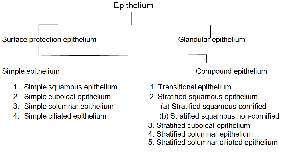
According to the functions, the epithelial tissues are mainly classified into the following two groups:
- Surface protection epithelium
- Glandular epithelium
Each group is again subdivided as follows:
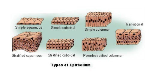
1. Simple Epithelium
The epithelial cells when arranged in one layer are known as simple epithelium. Simple epithelium is of the following types:
(a) Simple Squamous Epithelium
It is composed of single layer of flattened cells of different shapes, usually polygonal and of varying size. These cells fit together by their edges like the tiles of a mosaic pavement. The nucleus generally flattened but may be spheroidal.
This kind of tissue forms the lining of the alveoli of lungs, Bowman`s capsule, Henle`s loop of nephron, the inner lining of the heart (endocardium) and blood vessels(endothelium).
Functions
- It forms the outer surface of the body and is concerned in protecting the organism.
- It allows easy passage of gases by diffusion and fluid by filtration or dialysis process respectively.
(b) Simple Cuboidal Epithelium
- It is composed of single layer of cuboidal cells having same dimensions on each side. The cells adhere to one another by their lateral surfaces. The nucleus is spherical. On the free surface, this epithelium appears as mosaic of small, usually hexagonal polygons.
- Cuboidal epithelium is present in the germinal epithelium of ovary, on the thyroid follicles, in the excretory ducts of many glands, small terminal bronchioles and proximal convoluted tubules of nephrons.
Functions
- It acts as a protective layer in some parts of the body.
- In some part, it helps in secretion and storage.
- It forms germinal epithelium (ovary).
(c) Simple Columnar Epithelium
It is composed of single layer of tall (cylindrical) cells situated on the basement membrane. The height of the cell is more than the width. The nucleus is elongated or oval shaped and is situated nearer to the base. The free surfaces of some columnar cells are seen to be vertically striated. These striations look like brush border and known as microvilli. The free surfaces of the cells appear as small polygons. In some places (large intestine) the columnar cells are modified into flask-shaped mucus secreting cells, known as goblet cells.
It is found in the mucus layer of gastrointestinal tract, mucus membrane of the gall bladder, greater part of the male urethra, the vas deferens and duct of postrate, the large duct of salivary gland.
Functions
- The columnar epithelium is concerned with absorption and secretion.
(d) Simple Cilliated Epithelium
The cells of the epithelium are generally columner in shape but sometimes it may be cuboidal. The free surface has got 20-30 eyelashes (hair-like) processes. They are known as cilia. The border of the cell contains a raw of particles known as basal particles. From each basal particle, a cilium projects from free surface and longitudinal filaments called rootlets. It penetrates through the cell substance and converge to a point close to the fixed end.
It is distribute in the respiratory tract, uterus and fallopian tube, ventricles of brain and central canal of spinal cord, efferent tubules of testis.
Functions
The main function of the cilia is to move permanently so long as the cell is lining, By this movement ciliated ciliated epithelium maintains the following functions:
- Protection: In the fallopian tube it carries the ovum to the uterus.
- Circulation: The circulation of cerebrospinal fluid in ventricles of brain and central canal of spinal cord takes place with the help of ciliated epithelium.
You might also read: Neuron
2. Compound Epithelium
The epithelial cells when arranged in two or more layers on the basement membrane are called compound epithelium. The inner most layer of cells come in contact with the basement membrane, Compound epithelium is the following types:
(a)Transitional epithelium
This was originally supposed to represent transition, i.e., in between the stratified squamous non-cornified and stratified columnar epithelium. It contains three or four layers of cells. The cells of the most superficial layer are large quadrilateral flattened cells. They have a convex free surface round the depression on their under surfaces to fit on to the rounded ends of the second layer. The cells of second layer are pear-shaped and is known as pyriform cells. The next one or two layers consist of small polyhedral cells present in between the pointed ends of the pyriform cells.
This epithelium occurs in pelvis of the kidney, ureters, urinary bladder and upper part of the urethra.
Functions
- It acts as protective tissue and prevents re-absorption of excreted material(urine) into the body.
(b) Stratified Squamous Epithelium
It consists of several layers of cells which are different shapes. The more superficial layer is of flattened scale-like cells. The deeper layer of the cells varies from cuboidal to columnar. The cells remain adjacent to the basement membrane show considerable irregularity. According to the nature of the free surface, the stratified squamous epithelium is two types:
(i) Stratified squamous cornified epithelium: When the suoerficial cell layer of stratified squamous epithelial tissue is dry and undergo a transformation into a tough, resistant, dead horny nature due to deposition of Keratin is known as stratified squamous cornified epithelium.
It is distributed in skin, hair, nails, horns, enamel of teeth, etc.
Functions
Due to the presence of keratin, it performs the following functions: It is resistant to friction; It is relatively impervious to bacterial invasion; It acts as waterproof; It prevents drying up of underlying cells.
(ii) Stratified non-cornified epithelium: The superficial scale like cells are moist and is not keratinized and thus it is known as non-cornified epithelium.
It is distributed in vagina, female urethra, oesophagous, epiglottis, cornea, a part of conjunctiva, etc.
Functions
- It affords mechanical protection against friction and injury.
(c) Stratified Cubical Epithelium
It is composed of two or more layers of cubical cells. The superficial layers are smaller cubical than those of the basal layers. It is found only in the duct of sweat gland.
Functions
- It performs the functions of protection and secretion.
(d) Stratified Columnar Epithelium
It consists of several layer of cells. The deeper layer consists of small irregularly polygonal or fusiform cells. Only the cells of the superficial layer are of the tall columnar type.
It is found only in male urethra, in some excretory ducts, some parts of pharynx, epiglottis, anal mucous.
Functions
- It affords mechanical protection and helps in secretion.
(e) Stratified columnar ciliated epithelium
The cells are arranged as in the preceding type. The free surfaces of superficial columnar cells are provided with cilia. It is found only in the upper surface of the soft palate, some parts of larynx, etc.
Functions
- Protection and transport are the important functions of stratified columnar
Glandular Epithelium
The masses of secretory epithelial cells present in the different glands of the body are collectively known as glandular epithelium. During the embryonic development of the fetus, the secretory cell develops from the disintegrated cuboidal or columnar epithelium. The glandular epithelium is of the following types:
1. Based on the presence of number of cells in the glands
Glands are divided into two main groups, unicellular gland and multi-cellular glands.
Unicellular glands: In mammals, the only unicellular gland is mucous or goblet cell, which secret mucin. These goblet cells are found to remain scattered among the columnar epithelial cells of many mucous membrane.
Multicellular glands: The glands made up of many cells are known as multicellular gland. The secretion of these glands reach the free surface either directly or through excretory ducts.
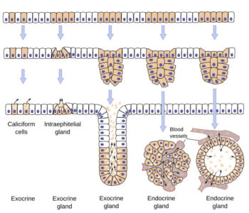
Figure: Showing different types of glands and their development. Image Credit-University of Vigo
2. According to mode of secretion
Glands may be divided into exocrine and endocrine types. The exocrine glands are also known as duct gland which secrets materials into ducts leading to a specific compartment or surface such as the lumen of gastrointestinal tract or the skin surface. The endocrine gland or ductless gland has no ducts and secretes material into the blood stream more precisely, into the extra cellular space around the gland cell from which the secretion diffuses into capillaries.
3. According to the structure of ducts and alveoli
Exocrine multicellular glands may be simple or compound depending on whether the ducts carrying the secretion to the surface are branched or un-branched.
(a) Simple exocrine gland
They consists of a secretary cells connected to the surface epithelium directly or by an unbranched duct. The simple glands of human are of the following types:
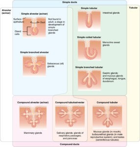
- Simple tubular gland: The terminal portion is straight such as intestinal glands.
- Simple coiled tubular gland: The terminal portion is a long coiled tubule which passes into a long excretory duct such as sweat gland.
- Simple branched tubular glands: The tubules of the terminal portion are slit fork like into two or more branches which sometimes coiled near ends such as glands of stomach, uterus, brunner glands of small intestine, etc.
- Simple branched acinous or saccular gland: If the terminal portion has the form of a spherical or elongated sac, the gland is called acinous or alveolar. If only one acinous is present with one excretory duct, it is a simple acinous gland such as the mucous gland of the frog`s skin but it is not found in mammals. If several acini are arranged alon a duct, it forms a single branched acinous gland such as sebaceous glands of the skin.
(b) Compound exocrine glands
A compound gland consists of many lobes. Each lobe is made up of many lobules which contain glandular units. Compound glands are of the following two types:
- Compound tubular gland: The terminal portions of the smallest lobules are more or less coiled and usually branching tubules such as glands of the oral cavity, some glands of the stomach, Brunner`s gland of the intestine, etc.
- Compound acinous or alveolar gland: The terminal portions are supposed to have the form of oval or spherical sacs e.g. most of the layers of exocrine glands such as glands of the oral cavity and respiratory passages and pancreas.
Figure: Types of Exocrine Glands- Exocrine glands are classified by their structure. Image Credit-Openstax.org
4. According to the nature of secretion
Compound exocrine glands are also occasionally subdivided into mucous, serous or mixed type.
5. According to the manner in which their secretary products are discharged, the glands may be divided into three types:
- Holocrine gland: In some glands, the entire secretory cells having formed accumulated secretory products within its cytoplasm, disintegrates and is discharged from the gland as the secretion. Such glands are sebaceous gland.
- Apocrine gland: The secretory products accumulate at the free surface. It then discharges its secretion by rupturing the free surface of the cell. After a recovery period, the cell again recharges to repeat the same cycle, such as mammary gland, apocrine sweat gland.
- Merocrine gland: The majority of glands are of the merocrine type whose secretory product is formed in and discharged from the cell without the loss of any cytoplasm. Such glands are salivary and pancreatic glands.

