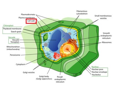
The cell wall is the semi-rigid, laminated, semi-permeable non-living outermost cellulose coat which is positioned next to the cell membrane of the plant cells, Fungi, Algae, Bacteria, and some Archaea. The protoplasm of the plant cells is separated from the external world by the cell wall which is entirely lacking in animal cells. Generally, the composition of the cell wall varies from organisms to organisms. The cell wall of the plant cell is composed of cellulose (Carbohydrates), bacterial cell wall contains sugar and amino acid polymer which is known as peptidoglycan while fungal cell wall is composed of chitin, glucans, and proteins. The cell wall performs lots of functions such as structure, protection, and support.

Molecular Structure of Plant Cell Wall
Formation of Cell wall
The cytoplasmic organelles like Endoplasmic reticulum, Golgi bodies, etc. take part in the formation of the cell wall. During cytokinesis process, small vesicles of endoplasmic reticulum migrate to the equator of the dividing cell and fused with one another to form barrel-shaped discontinuous cell plate or phragmoplast. Golgi bodies synthesize all the polysaccharides as pectin, hemicelluloses of α-cellulose, microfibril of the cell wall of plant cells. All these polysaccharides along with cell plate help in the cell wall formation.
Chemical Composition of Cell Wall
1. Matrix
2. Microfibrils:
- Cellulose: 10-15%.
3. Other Ingredients
- Lignin
- Cutin
- Suberin
- Silica (silicon dioxide)
- Minerals: Iron (Fe), Calcium (Ca), Carbonate (CO3), Waxes, Tannins, Resins, Gum, etc.

Image showing cell wall and major chemical composition
Structure of Cell Wall
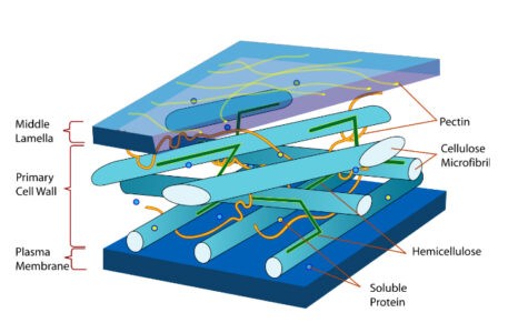
The plant cell wall is multi-layered and it is complex in nature. There are three distinct layers in the cell wall. From the outermost layer of the cell wall, these are the middle lamella, the primary cell wall, and the secondary cell wall. Occasionally tertiary cell wall may also be present. All plant cells contain a middle lamella and primary cell wall but not all have a secondary cell wall.
1. Middle Lamella
It is comparatively thin viscous and jelly-like intercellular cementing material which is present in between the primary cell walls of two adjacent cells. It is first formed after cell division (Cytokinesis). It is mainly composed of pectin (polysaccharides), calcium and magnesium, etc. Pectin acts as intercellular cement which helps in cell adhesion to bind the adjacent cells together.
2. Primary Cell Wall
It is the outermost thin elastic permeable membrane which is situated between the middle lamella and plasma membrane in growing plant cells. It is formed during the early stages of growth and development. It is composed of cellulose, hemicelluloses, pectin, and lignin, etc. It is 1-3 µm thick. The primary cell wall provides the flexibility and strength which is essential for cell growth.
Secondary Cell wall
It is thick permeable cell wall which is present in between the primary cell wall and plasma membrane in some plant cells. As the cell matures and differentiates, the secondary cell wall is deposited. Thus, further cell expansion ceases. The secondary cell wall is composed of organic compounds like cellulose, hemicelluloses, lignin, suberin, cutin, wax, etc. and inorganic salts like calcium carbonate, oxalate, silica, etc. It is about 6-10 µm thick. The secondary cell wall commonly has three layers, such as a thin outer layer, thick middle layer, and a thin inner layer. This rigid layer makes strength, gives support to the cell and helps in water conductivity in plant vascular tissue cells.
In some cells, a very thin tertiary cell wall is formed on the inner surface of the secondary cell wall. It is made up of xylan instead of cellulose.
Ultra-structure of Cell Wall
Electron microscope revealed that the cell wall is composed of two main parts such as microfibrils and matrix.
Microfibrils: Each microfibril is 0.5 µm in width and about 250 Å in diameter. Each microfibril contains a bundle of micelles of elementary fibrils. Each micelle is about 100 Å in diameter and contains about 100 cellulose chains. Cellulose is a polysaccharide made of glucose arranged side by side.
Matrix: Matrix consists of largely of polysaccharide. In it, the microfibril of cellulose and other polysaccharides are embedded.
Plasmodesmata
The cell wall does not form a continuous layer but has many small openings or pores. In the cell wall at many places, the cellulose layers are absent and at these places, small pits are formed. These places are called primary pit fields. These pits are always opposite in between the walls of adjacent cells. In such a case, the middle lamella is called a pit membrane. There are many minute pores in the middle lamella. The cytoplasm of one cell communicates with the cytoplasm of adjacent cells through these pores together by microscopic fibrils. Each fibril is called plasmodesma and the group of these fibrils is called plasmodesmata (singular-plasmodesma).

Plasmadesma
Thickening of the Cell Wall
Secondary wall is thicker and more rigid than the primary cell wall due to lignin deposition. These depositions occur in such a way that various many peculiar patterns of ornamentation are formed on the cell wall such as:
1. Annular: The materials of the secondary cell wall are deposited inside the primary cell wall. This deposition forms the ring like thickening at regular intervals and the rest of the wall is thin. This type of structure is present in the protoxylem vessels.
2. Spiral: The matrix of the secondary cell wall is deposited inside the primary cell wall. This deposition occurs in such a way that forms the spiral or helical thickening. This is also found in the protoxylem vessels.
3. Scalariform: The secondary wall matters are deposited irregularly in such a way that forms a staircase-like structure.
4. Reticular: The matters of the secondary wall are deposited irregularly on the primary cell in such a way that looks network-like structure.
5. Pitted: The matters of the secondary cell wall are deposited uniformly all over the primary cell wall except certain areas. These areas look like cavities which are known as pits.

Different Types of Cell Wall Thickening
Pits
Generally, pits occur in pair, called pit pairs, i.e., when a pit is formed on the cell a complimentary pit will be formed just on the opposite side of the adjacent cell. Pits are usually found in non-living cells like tracheids and fibers. Pits are of two types: Simple and branched pits.
Simple pits: They have no projecting margin or border around the aperture. Simple pits are found in sclerids, xylem parenchyma, ordinary parenchyma cells, vessels, etc.
Branched pits: These types of pits are provided with projecting margin or border. They consist of (i) pit chamber which is the area within the pit and (ii) Pit aperture which is the aperture through which the pit chamber opens outside. (iii) Pit membrane– adjacent pits are separated by the middle lamella and the primary cell wall together form pit membrane. It may have a thickening area, called the torus which is formed by the circular deposition of microfibrils.
Functions of the Cell wall
- It provides protection against plant viruses and other pathogens;
- It gives mechanical support to the plant cells due to their semi-rigid exoskeleton.
- It provides rigidity, strength, and flexibility to the cell due to their thickening wall.
- It provides and maintains the structural integrity of the cell.
- The cell wall contains Cutin and suberin which helps to reduce or prevent water loss through transpiration.
- The cell wall acts as storage to store carbohydrates which are used in plant growth.
- The cell wall helps to the interchange of different substances. In this case, water, salt or other substances like protein can easily pass in or out through the cell wall due to their permeable characteristics.
- It also provides tensile strength and limited plasticity which help to prevent rupture of the cell from the turgor pressure.
- It transfers signals to the cell during cell division to control the direction of cell growth.
- Different physiological and biochemical activities occur in the cell wall which contributes cell communication with one another via plasmodesmata.
- The cell wall helps in cell expansion.
- The cell wall does some enzymatic activities which is connected with metabolism.
- Sieve tubes, tracheids, and vessels are present in the cell wall which helps in the long distance transportation of different substances.
- The cell wall plays a big role to form a framework for the cell to prevent overexpansion.
Also read: Plasma Membrane: Structure and Functions

