Introduction
The system through which blood circulates in different organs and parts of the body is called Blood Circulatory System. The presence of well-developed circulatory system can be observed in almost all animals with few exceptions. Fish have a closed type of blood circulatory system. Food, oxygen and waste products are transported from one part of the body to another through the blood flowing in such circulatory system.
The circulatory system is actively involved in controlling the metabolism of food, coordinating the various organs and systems of the body, preserving, repairing, and destroying various pathogens. Although the circulatory system has special features compared to other organs, its structure is equally common. The circulatory system of fish consists of blood, blood vessels (arteries and veins) and the heart.
Permeable membranes exist in most areas of the fish body. For this purpose, water is exchanged through the gills, and in addition to the gases dissolved in the gills, the exchange of some nitrogenous wastes and minerals is carried out. The entrance and return of blood from the body of the fish to the gills, except for the lung fish, is accomplished by a single circulation. In this case, the heart exchanges blood with low concentration oxygen and high concentration carbon dioxide.
The blood volume of higher bony fish (teleost) ranges from 1.5% to 3% of total body weight. In mammals, however, the amount of blood is 6% or more of body weight. Spiny dog fish (Squalus acanthias) has a blood volume of 5% of body weight. Plasma or blood cells of fish are produced in greater quantities in different organs or systems than in mammals.
A notable feature of the circulatory system of fish is that there are a significant number of capillary or sinusoidal systems in the arterial or venous flow of blood. The special system created as a result of such capillary arrangement is called portal system. Such systems are found in the gills, liver (hepatic portal system) and kidneys (renal portal system). There is also another capillary resembling vessels in the rete mirabile in one part of the swimbladder of Physoclystous fish. The arrangement of the chloride glands in the eyes of the teleost is similar.
Some fast-moving fish such as mackerel sharks (Lamnidae), tuna, mackerel (Scombridae) have other organs such as special capillary blood vessel in muscles. As a result of this system, the exchange of gas between blood and tissues is done more efficiently.
Blood
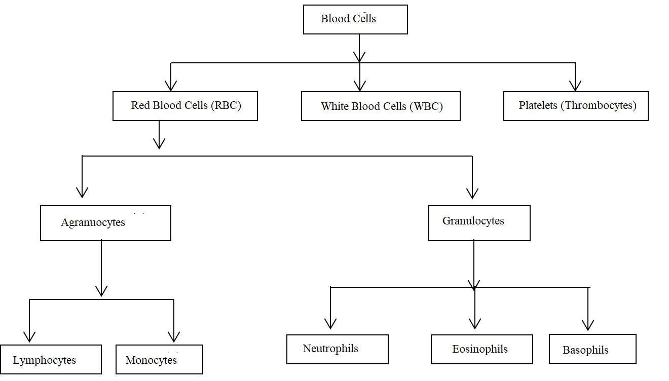
The blood of fish is the same connective tissue as other vertebrates. The liquid part is called plasma and the solid part is called blood cells and other substances which are in the liquid part. It includes the following blood cells: red blood cells (erythrocytes or RBCs), white blood cells (leukocytes or WBCs) and platelet (thrombocytes). Red blood cells are red in color because they carry a type of red pigment called hemoglobin. It plays an important role in the transport of oxygen in the blood.
Not all fish have red blood cells and hemoglobin. Some Antarctic fish (Chaenichthyidae, icefish or white crocodile fish) have colorless blood because they do not have erythrocytes. The blood of the small eel (Leptocephalus larvae) is also colorless. Blood pigment of Lamprey (Petromyzon) is not like the hemoglobin of other vertebrates.
Plasma
The clear liquid that is obtained by separating blood cells from blood is called plasma. In the broadest sense, if blood is collected in a bottle with anticoagulants, the blood will not clot, and in this case, if the blood is centrifuged, the blood cells will be separated and stored as sediment, then the remaining liquid is called plasma.
If the blood is collected in a bottle without anticoagulant, the blood will clot and in this case, if it is centrifuged then the liquid part is called serum. In fact, serum loses the blood clotting component, called prothrombin and fibrinogen, but plasma carries the proteineous blood clotting component.
Plasma contains various protein components (fibrinogen, globulin, albumin, etc.), dissolved minerals (Na +, K +, Ca ++, Mg ++, Cl–, HCO3–, PO4—, SO4—), absorbed component as a result of digestion (glucose, Fatty acids, amino acids), tissue waste products (urea, uric acid, creatine, creatinine, ammonium salts), special secretions (hormones and enzymes), antibodies and dissolved gases (oxygen, carbon nitrogen). The sedimentation co-efficiency of major plasma proteins varies from species to species.
The electrolyte (ion) per liter of blood in cod fish (Gadus callarius) is 180 ml of sodium (Na +), 4.9 ml of potassium (K +), 3.8 ml of magnesium (Mg ++), 5.0 ml of calcium (Ca++), 5.3 ml of chloride (Cl–), 3.1 ml of phosphate (PO4—). Concentrations of sodium and chloride are generally lower in freshwater teleost.
Sharks (Squaliformes) have high concentrations of Mg ++ in their blood. However, its blood is mildly alkaline than the blood of higher bony fish (Actinopterygii). The dissolved material in solution indicates the freezing point which can also be measured by osmotic pressure. As the osmotic pressure of the blood increases, water spreads from the permeable membrane to the low density solution.
In freshwater bony fish, the freezing point of plasma is 0.50C. For some freshwater sharks and other fish (elasmobranks) it is 1.00C. For marine bony fish, the value is 0.6-1.00 C. The maximum value in marine elasmobranch is 2.170C. The freezing point of seawater is 2.080C.
Fish have lower plasma proteins than higher vertebrates. The major plasma proteins in fish are albumin (regulating osmotic pressure), lipoprotein (transporting lipids), globulin (binding to hemi part), ceruloplasmin (binding to copper), fibrinogen (helping to blood clots) and iodi-uroforin ( only found in fish, adding inorganic iodine).
The concentration of plasma protein in fish is 2-6 g / liter. The presence of low levels of fibrinogen and prothrombin-like proteins is not associated with rapid blood clotting. Rainbow trout (Salmo gairdneri) can survive above 00 C. At low temperatures, the blood of this fish clots. Because serum of Antarctic fish contains glycoproteins, they can survive at -1.90C. The ratio of albumin and threonine in this protein is 2: 1. Its molecular weight is 2600-33000.
Thyroid-binding proteins such as T3 and T4 are found in the blood plasma of fish. In cyprinid species, it adds vitalogenin. It also contains a variety of enzymes such as CPK, alkaline phosphatase (Alk Pase), SGOT, SGPT, LDH, lipase and carbonic anhydrase and their co-enzymes.
If the serum of some teleosts, especially Anguilla, some catfish (Siluridae), and the tuna (Thunnis) push into the blood of mammals then it shows poisoning reaction.
Types of Blood Corpuscles
The types of blood cells are mentioned in the following diagram:
1. Red Blood Corpuscles/Erythrocytes
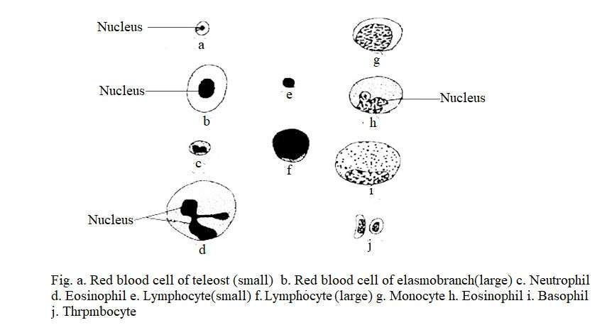
Most fish have red blood cells with round or rectangular nuclei that are located in the center of the cell and are yellowish red in color. Its numbers vary depending on the species, age, season and environmental influences. Its size is large in Elasmobranch and small in teleost. In estuary species such as Fundulus, it is smaller in size than freshwater species.
Red blood cells of deep sea teleosts are larger in size than common teleosts. In species like Clarias batrachus, Notopterus notopterus, Colisa fasciatus, Tor tor, etc., its structure is usually round but in Labeo rohita and Labeo calbasu it is oval in shape.
Some species of Antarctic fish that live in oxygen-rich areas at low and cool temperatures do not have red blood cells. In addition, leptocephalus larvae of eel fish (Anguilla) and some deep-sea fish do not have red blood cells. Their gas exchange occurs through diffusion. The red blood cells of the fish are oval, small and 6 microns in diameter, but in many fish, especially Wrassus (Crenilabrus), the red blood cells are more than 8 microns in diameter. In Protopterus, it is 36 microns of diameter.
The number of red blood cells per cubic mm of blood in fish is 20,000-3,000,000. Inactive fish have a lower number of red blood cells than active fish.
2. White Blood Corpuscle
There has been considerable research on the white blood cells of fish, so there is no difference in their classification. The number of per cubic mm in the blood of fish is 20,000-150,000. It may be granulocyte or agranulocyte but the number of granulocytes is higher. Granulocytes can be further divided into eosinophils, basophils and neutrophils based on their staining capacity.
Neutrophils and eosinophils have phagocytic properties. Agranular white blood cells are lymphocytes and monocytes. Monocytes produce antibodies. Basophil granulocytes are found in some species, but no function of it has been reported.
A. Agranulocytes
(a) Lymphocytes: Different types of lymphocytes are found in the blood of fish. Their nuclei are round or oval in shape. Lymphocytes are 80-90% of the total white blood cells. It contains a lot of chromatin. Like mammals, freshwater and saltwater fish also have large and small lymphocytes. Large cells contain large amounts of cytoplasm. They do not have granules in their cytoplasm. The main function of lymphocytes is to increase immunity by making antibodies.
(b) Monocytes: Monocytes account for small quantities of white blood cells. However, some fish do not have monocytes. They are thought to originate from the kidneys and are visible when an unwanted object enters the bloodstream. Its cytoplasm is light blue or purple in color. The nucleus is basically large and has a variety of structures. Its main function is to destroy pathogens in the process of phagocytosis.
B. Granulocytes
(a) Nutrophil: Most of the white blood cells in fish are neutrophils. Neutrophils are 5-9% of the total white blood cells in Solvelinus fontinalis and 25% in brown trout. They are named based on the staining capacity of the cytoplasm. Their nuclei are multi-lobed but some fish have neutrophils with bi-lobe nuclei.
In marginal blood smears, the cytoplasm contains pink, red or purple granules. Their nuclei look like human kidneys. Neutrophils react positively with peroxidase and Sudan Black. Neutrophils are active phagocytes. It protects the tissues from inflammation or injury.
(b) Eosinophils: Eosinophils are usually round and its cytoplasm is granular. In acidic solution, it shows dark pink orange or orange red color. Their nuclei are lobed and show dark orange to purple in color.
3.Thrombocyte
This is also called platelet. They are round, oval or spindle shaped. In mammals, however, the platelets are disc shaped. Fish blood contains platelets which is about half of the total leukocytes.
Herring fish contain 72.2% platelets of red blood cells and only 0.7% of platelets in teleost. Their cytoplasm is granular, the center is more alkaline and its circumference is dull and homogeneous type. In alkaline solution, their cytoplasm shows pink or red color. They help in blood clotting.
Origin of Blood Corpuscle
The process of forming blood cells and blood plasma is called hemopoiesis. In the early embryonic stage, blood cells are produced from the wall of the blood vessel. Red blood cells and white blood cells originate from lymphoid hemoblasts or hemocytoblasts and enter the bloodstream to mature.
In fish, spleen and lymph nodes are involved in the production of blood cells. In chondrichthyes, red blood cells originate from the granulopoietic tissue, the leidig organs, the epigonal organs, and rarely the kidneys. The leidig organ is made up of white tissues and acts like a bone marrow tissues. Such tissues are found in the esophagus but their main source is the spleen. In all these fish, if the spleen is removed, the leidig organs take part in the production of red blood cells.
In teleost, red blood cells and granulocytes originate from the kidneys (pronephros) and spleen. Their spleen has a red cortex on the outside and a medulla with white pulp on the inside. Red blood cells are produced from cortical region of the spleen while lymphocytes and some granulocytes are produced from the medullary region.
The intestinal spiral valves of chondrichthyes and Dipnoi also produce different types of white blood cells. In higher bony fish (Actinopterygii), red blood cells are destroyed in the spleen. The technique of destroying blood cells in jawless fish (Agnatha), Basking shark and Rays is not known.
Thrombocytes originate from the mesonephric kidney of fish, granulocytes originate from the submucosa, liver, gonads, and mesonephric kidney of the digestive tract.
In sharks, rays, and chimaera (chondrichthyes), white blood cells with connective tissues are seen below the mucous membrane of the esophagous. In sturgeon (Acipenser), paddle fish (Polyodon) and South American lung fish, the reddish-brown lobular spongy tissue around the heart produces lymphocytes and granulocytes.
Skull bones of some sharks (Squaliformes), chimaeras (Chimaeridae), Gar (Lepisosteus) and cranial cartilage of Bowfin (Amia) can produce all types of blood cells.
Function of Blood
Like other vertebrates, the mixed cellular components of blood plasma is found in fish. It consists of a type of one kind of connective tissue and a non-newtonian fluid. Blood flows throughout the body through the cardiovascular system. It is mainly caused by contraction of the heart muscle. Blood has different functions. The functions of blood are given below:
1. Respiration: Blood plays an important role in transporting dissolved oxygen(DO) from water to the gills (respiratory changes) and carbon dioxide(CO2) from the tissues to the gills.
2. Nutrition: The blood carries various nutrients such as glucose, amino acids, fatty acids, vitamins and electrolytes, and secondary elements from the alimentary canal to the tissues.
3. Excreation: The waste products produced from blood metabolism such as urea, uric acid, creatine etc. are carried away from the cells. All fish have trimethyl amine oxide in their blood, but its concentration is highest in marine elasmobranch.
Creatine is a type of amino acid that is produced by the metabolism of glycine, arginine, methionine. The amount of creatine in the blood plasma is 10-60 grams and it is excreted through the kidneys.
4. Homeostasis of Water and Electrolyte Concentration: The exchange of electrolytes and other molecules takes place through the blood. The level of glucose in the blood of fish is considered to be a sensitive physiological indicator in most cases and there is no discrepancy in the level of glucose in the blood of fish.
5. Hormones: There are different types of controlling agents in the blood such as hormones and cellular or humoral agents (antibodies). All these elements are present in different concentrations in the blood which are regulated by the feedback loop and change the concentration and make the necessary components of different organs through the synthesis of hormones and enzymes.
Heart: Structure and Functions
The heart is a special pump device with a valve in the circulatory system. In case of fish, the heart is a folded tube that contains three or four enlarged areas. The blood brought through the veins travels from the heart to the gills through the ventral aorta and the heart always contains carbon dioxide (CO2) or unrefined blood. This is why the heart of a fish is called the venous or branchial heart.
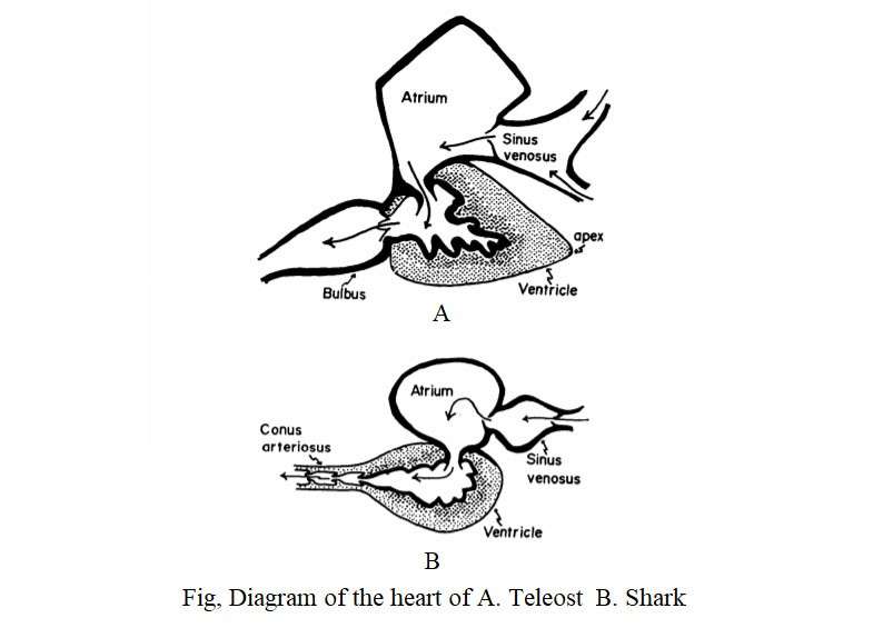
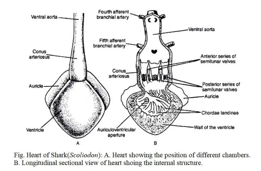
Blood passes through the aortic arch on the front and enters the gills to exchange gaseous components through the heart. In most fish, the heart is located immediately after the gills. In the case of the teleost, the heart is located at the front side of the body rather than at Elasmobranch. The heart is primitive type in Elasmobranch among the fish. It is located in the pericardial cavity and consists of the sinus venosus, atrium, ventricle and well-developed contractile conus.
Some researchers consider the atrium and ventricles to be the chambers of the heart. Some researchers also consider the sinus venosus and conus arteriosus to be the chambers of the heart. In the case of fish, there is some controversy over Conus arteriosus and Bulbus aorta. The fourth chamber of the elasmobranch is known as the conus arteriosus. In Teleost, however, it is known as Bulbus arteriosus.
The difference between conus arteriosus and bulbus arteriosus is that conus arteriosus has ventricular like heart-muscle and innumerable valves are continuously arranged in it, but bulbus arteriosus consists only of smooth muscle fibers and elastic tissue. (Boas 1980; Smith 1918; Danforth 1912; Parson 1929; Karandikar and Thakur 1954).
According to Torrey (1971), a teleostian fish (Cyprinus carpio) has both conus and bulbus arteriosus. According to Kumar (1974) and Santer (1977), the teleost has only bulbus arteriosus. On the other hand, Elasmobranch and Agnatha have conus arteriosus instead of bulbus arteriosus.
The heart has sino-auricular and sino-ventricular openings that are controlled by a dual valves. The conus has six rows of valves. Muscular and contractile conus are considered to be of primitive nature. It is found in some lower teleosts such as Acipenser, Polypterus and Lepidosteus.
In addition to conus, bulbus arteriosus exists in the Amia. It originates from the fibrous wall of an uncontrollable region. This type of secondary condition is seen in some lower teleost (Clupeiformes). Albula, Tarpon and Megalops have distinct conus and rows of transverse valves. The size and weight of the heart varies with the body weight of the fish. The structure of the heart of different fish is described below:
Heart of Cyclostomes
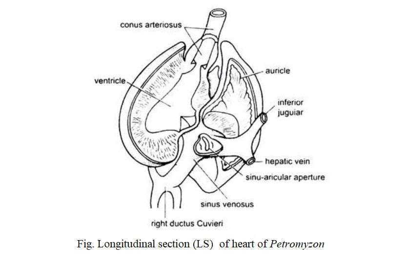
Lamprey’s (Petromyzon) heart is like the English letter ‘S’. It is formed by folding the posterior side of the gill and the sub-intestinal vessels. The larval heart develops as a straight duct. Later, this duct becomes longer and takes the shape of ‘S’ in a limited space. The heart consists of the sinus venosus, an atrium and a ventricle, and the conus arteriosus and is covered by the pericardium. A cartilage plate holds the pericardium. The sinus venosus is a thin-walled chamber that is exposed through a sinus-auricular opening to a thin-walled atrium at the top. The atrium is again connected to the thick-walled ventricle through the auricle-ventricular opening.
Heart of Cartilaginous (shark) Fishes
Their heart is a curved muscular duct that consists of the receiving region and the transmitting region. The receiving region consists of a sinus venosus and dorsally located an atrium while anterior portion contains a ventricle and a conus arteriosus. The heart is covered by a membrane called the pericardium. The dorsal part of the pericardium is made up of basibranchial cartilage. The heart is located between the two rows of gill pouches on the ventral side of the body of the fish.
Heart of Bony Fishes
(a) Heart of Tor tor
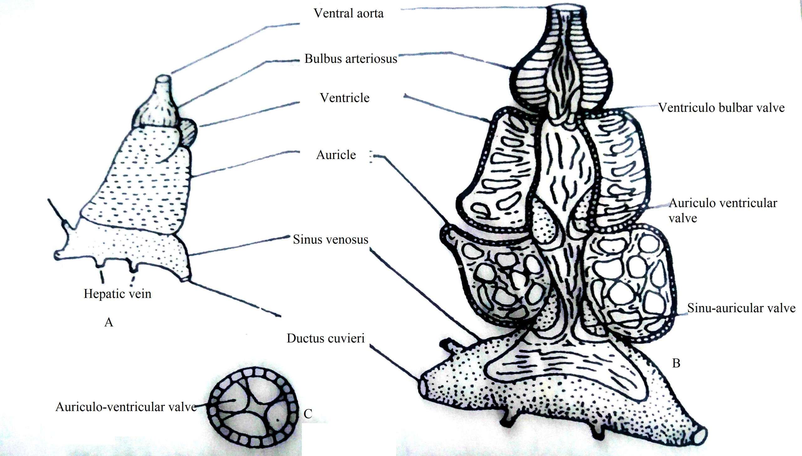
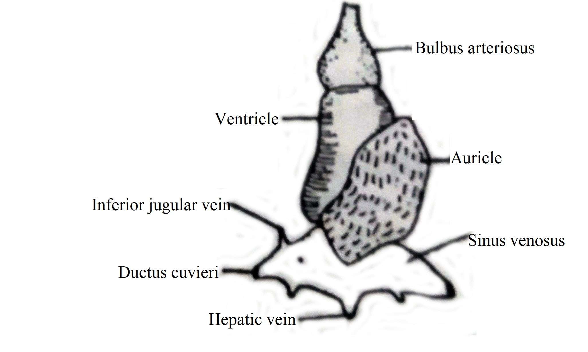
The heart is located at the tip of the septum transversum in the pericardium sac. It consists of sinus venosus, atrium, ventricle and bulbus arteriosus. The sinus venus is a smooth-walled chamber that receives blood supply through the joint Ductus cuvieri, the joint hepatic vein, a posterior cardinal, and an inferior jugular vein. The openings of these blood vessels have no valves. The sinus venosus is exposed to the atrium through the sinus-auricular opening. In this opening, a pair of membranous semilunar valves are present. Each valve has a long wing pointed towards the front of the atrium.
Heart of Tor tor A. Internal structure of heart B. Cross section of heart
The atrium encloses the ventricle dorsally and is relatively large in size with an irregular outer surface. It is orange and sponge-like soft and has a narrow cavity extending to the ventricles. The spongy wall of the atrium has numerous spaces or cavities covered by muscle fibers extending in different directions. The atrium-ventricular opening contains two pairs of semilunar valves of approximately equal shape. Each valve has a short wing adjacent to the atrium wall and a long wing adjacent to the ventricular wall, but the extended part of the valves points towards the atrium.
The ventricle is a superior muscular chamber with a thick wall and a narrow cavity. It is associated with bulbus arteriosus through ventricular-bulbar opening. There are a pair of semilunar valves in this opening. Each valve has a short wing adjacent to the ventricular wall and a long wing adjacent to the wall of the bulb so that the wings cross each other. The valves are hanging in the ventricular cavity. The wall of the bulbus is thin and has a narrow hole in it. In its cavity, a thin ribbon-like innumerable trabeculae pass parallelly. The bulbus extend into the ventral aorta anteriorly.
(b) Heart of Other Teleosts
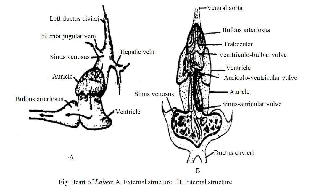
The heart of cyprinids such as Labeo rohita, Cirrhina mrigala, Catla catla and Schizothorax has the same general structure as Tor tor. Lebeo rohita, Cirrhina mrigala, and Catla catla have large sinuses and a pair of lateral appendages (Singh 1960). In the first two species, it is spongy and fibrous.
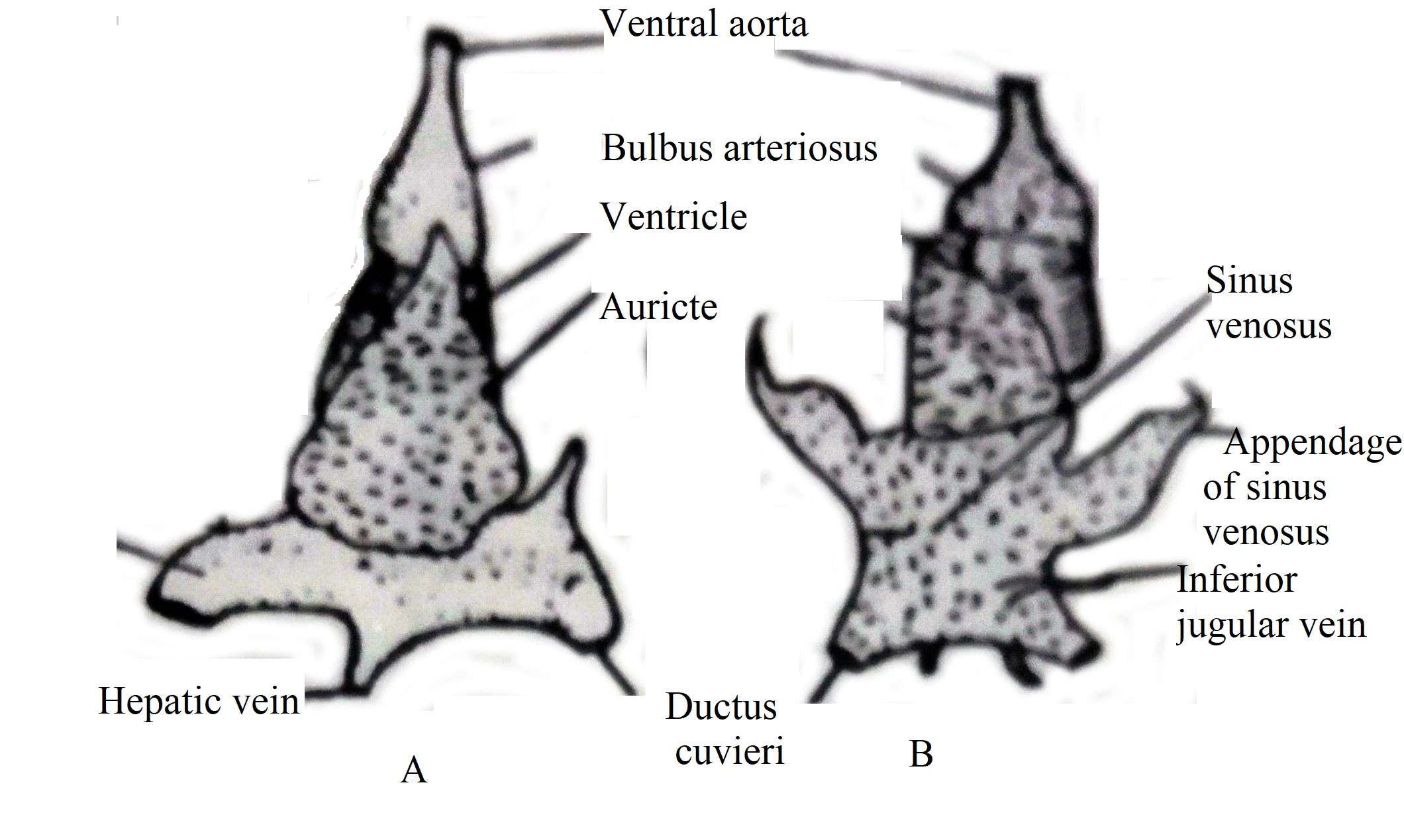
In Clarias batrachus, Mystus aor, Wallago attu, the sinus venosus is a thin-walled chamber in which a pair of membranous sinus-auricular valves are located obliquely along the long axis of the heart. One end of the dorsal valve extends to the front and reaches the atrium cavity and is attached to it.
Fig. Showing Heart of A. Wallago attu B. Catla catla
The atrium is structurally spongy type, looking like a beehive. The opening of the atrium ventricle has four valves, two of which are well-developed, while the other two are small, not so important. The ventricle has advanced muscular and two ventriculo-bulbar valves which are semilunar-like in shape. In case of Channa striatus, sinus venosus is small and there is no sinus-auricular valve.
Fig. Showing Heart of Clarias batrachus
Notopterus notopterus has 5-7 nodular valves in the sinus-auricular opening and Chitala chitala has 8-10 valves. Two of the 4 auricular-ventricular valves are small in size. Chitala chitala has a muscular conus arteriosus between the ventricles and the bulbus. In case of Notopterus, the ventricular bulbar valve is like a ribbon with a strange structure and divides the bulbous cavity into three chambers by a pair of vertical septum.
Working of the Heart
The venous blood travels to the heart, reaches the sinuses applying pressure to the semilunar valve, and reaches the atrium. During this time, the pockets of the valves are filled with blood and the pressure created by the contraction of the atria causes the valves to swell and obstruct the flow of blood from each other.
Due to the pressure of the four auricular-ventricular valves, the blood reaches the ventricle from the atrium and as soon as possible the ventricular cavity is filled with blood. During this time, the valves receive blood. So the valves swell and close the openings from being firmly attached to each other. As a result, the reverse flow of blood is obstructed. The blood then enters the bulbus by applying pressure to the ventriculo-bulbar valve. Inside the bulbus, the blood pressure rises again, causing the valves to swell and close the passageway, obstructing the retrograde flow of blood, causing the blood to flow forward through the ventral aorta.
Cardio-vuscular Control
Fish control the cardio-vascular system in two ways, viz
(1) Aneural and
(2) Neural mechanisms.
Aneural cardio-vascular control is accomplished through the direct response of the heart muscle to changes in temperature and the secretion of various glands and changes in blood volume. Temperature acts as an anural regulator due to the direct action of the myocardium on the pacemaker. In some species, an increase in temperature increases the heart rate, resulting in higher cardiac energy. By increasing blood flow, it is able to supply more oxygen to the body. As a result, higher metabolic rate is possible in warm water. Anural control also occurs under the influence of certain hormones such as epinephrine stimulates heart rate.
Neural control techniques occur through the tenth carotid nerve (vegus). The heart of these fish is nerved by a branch of the vegus nerve. Stimulation of the vegus nerve reduces the heart rate in the elasmobranch and teleosts. Different types of stimuli such as flashing light, sudden movement of an object, touch or mechanical vibration reduce the heart rate in fishes. In responding to environmental or other changes, fish face some problems during maintaining their blood circulation balance.
Arterial System of Lamprey
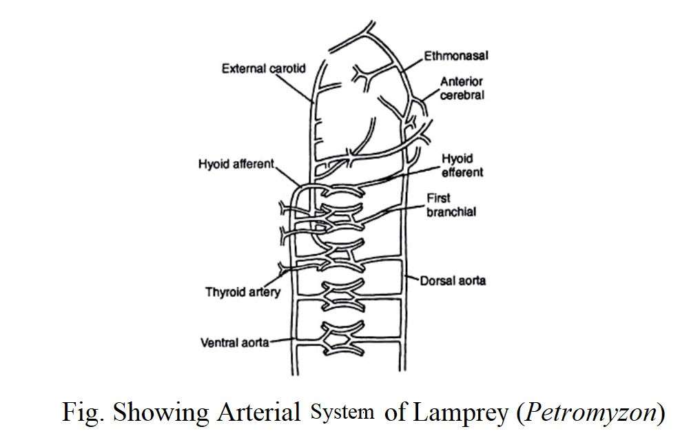
From the ventricle, a large ventral aorta emerges and moves forward through the gill pouches. The base of the ventral aorta is slightly swollen. Some researchers have named this swollen part as the bulbous arteriosus. Eight afferent branchial arteries from the ventral aorta enter into the gill pouches. The afferent branchial arteries divide into capillaries in the gills. Blood is collected from the gills by eight efferent branchial arteries.
Each of the afferent and efferent branchial arteries supplies blood to the posterior hemibranch of a gill pouch and the anterior hemibranch of the next. Each efferent branchial artery carry oxygenated blood from the gill pouch to the paired dorsal aortae. This paired dorsal aortae run backwards and joins to form a single median dorsal aorta. From this dorsal aorta, segmental arteries arise which enters into the myotomes. The segmental artery contains scattered chromafin cells that represent scattered adrenal medulla. Its secretion is similar to that of mammalian adrenalin.
Special arteries are produced from the unpaired dorsal aorta and supply blood to the intestines, kidneys and gonads. With the exception of the efferent branchial and renal arteries, most other arteries have valves at their origin. These valves play an important role in lowering blood pressure in most arteries. Blood flows forward through the ventral aorta and backwards through the paired and unpaired dorsal aortae.
Venous System of Lamprey
Their venous system consists of a complex network of true veins and sinus venouses. Blood is transported from the caudal region through a large caudal vein. This vein divides into two posterior cardinal veins just at the entrance into the abdominal cavity. Cardinal veins collect blood from the kidneys, gonads, and myotomes and ultimately opens to the heart by a single ductus cuvieri on the right side.
The left ductus caviary does not remain in adulthood. Although their presence can be noticed in the larval stage. Blood enters to the heart from the anterior region of the body through a pair of anterior cardinal veins. In addition to these anterior cardinal veins, a large median inferior jugular vein carries blood from the musculature of the buccal funnel and gill pouches. There are no renal portal veins in the lamprey. However, a hepatic portal vein collects blood from the intestine and enters into the liver through a contractile portal heart. A very simple type of portal system exists in the lamprey that connects the hypothalamus with the pituitary.
Blood from the liver enters into the heart through hepatic veins. In addition to the veins, special network of the venous sinus exists, especially in the head region. The branchial sinus is a very important sinus and consists of three longitudinal channels, namely:
(1) The ventral branchial sinus or the ventral jugular sinus;
(2) Inferior branchial sinus which is located below the gill pouches;
(3) Superior branchial sinus which is located above the gill pouches;
All these branchial sinuses are interconnected to each other by gill bars.
Arterial System of Cartilaginous Fish (Scoliodon)
The circulatory system of cartilaginous fish such as Scoliodon is composed of blood, heart, arterial system and the venous system. In Scoliodon, there are two distinct arteries in the arterial system, namely-
- Afferent branchial arteries; and
- Efferent branchial arteries.
The arterial system of Scoliodon is briefly described below:
1. Afferent Branchial Arteries of Scoliodon
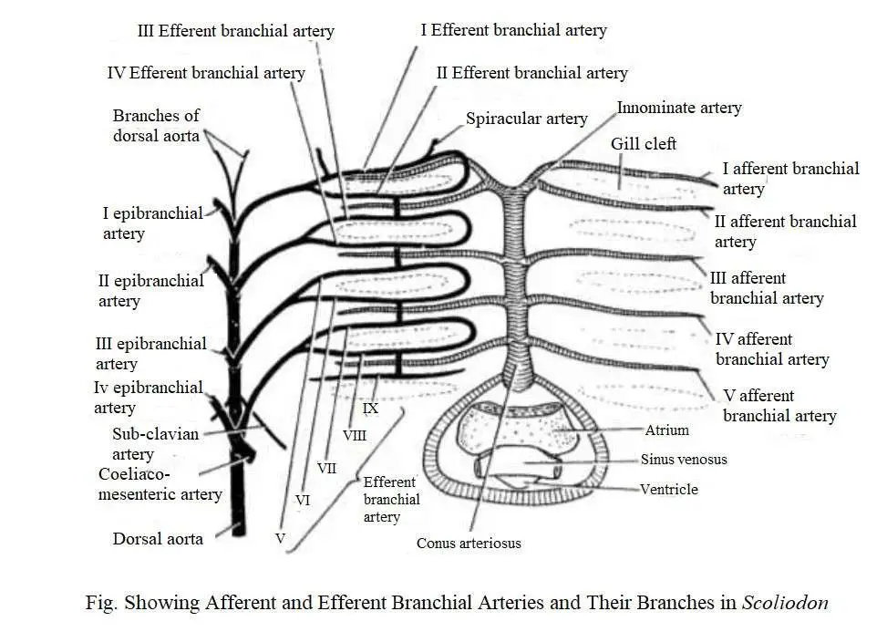
Afferent Branchial Arteries begin from the ventral aorta and carries oxygen-free blood to the gills for oxygenation. The ventral aorta is situated on the ventral surface of the pharynx. It extends up to the posterior boundary or hyoid arch. The ventral aorta is divided into two branches called the innominate arteries, each of which re-divides into two branches to form the 1st and 2nd afferent branchial arteries. The 3rd, 4th and 5th afferent branchial arteries originate from the ventral aorta.
Each afferent artery originates from a ventral aorta through an independent opening except for the 1st and 2nd afferent branchial arteries which are exposed to the same common opening.
2. Efferent Branchial Arteries of Scoliodon
Efferent Branchial Arteries arise from the gills and carries oxygenated blood to different parts of the body. The efferent branchial artery divides into capillary blood vessels in the gills. Blood is collected from the gills by efferent branchial arteries.
In Scoliodon, there are 9 pairs of efferent bronchial arteries that are evenly distributed on each side. The first 8 arteries form a series of four complete loops around the first four gill slits.
The 9th efferent branchial artery collects blood from the hemibranch of the 5th gill pouch and from where the blood is poured into the 4th loop. In addition, the shorter longitudinal connector connects the four loops. These are re-connected with each other by a network of longitudinal commissural blood vessels called the lateral hypobranchial chain.
An epibranchial artery originates from each efferent branchial loop. The four pairs of epibranchial arteries join along the mid-dorsal line to form the dorsal aorta. The 9th efferent branchial artery has no epibranchial branch. However, it joins with the 8th efferent branchial artery.
Anterior Arteries
The head region receives blood supply from the 1st efferent branchial artery and partly from the proximal end of the dorsal aorta. The following arteries originate from the 1st efferent branchial artery (hyoidian efferent), viz.
(a) external carotid
(b) afferent spiracular
(c) hyoidean epibranchial which gets blood from a branch of dorsal aorta.
The external artery receives blood from the first collecting loop and subsequently divides to produce a ventral mandibular artery and a superficial hyoid artery.
The ventral mandibular artery produces branches to the muscles of the lower jaw and the superficial hyoid artery which supplies blood to the 2nd ventral contractile muscle, the skin and subcutaneous tissue below the hyoid arch.
The afferent spiracular artery originates from the medial space of the hyoidian efferent artery and enters the cranial cavity as it progresses forward as the spiracular epibranchial artery. Just before its entry to the cranial cavity it sends large ophthalmic arteries to the eye ball.
As the spiracular epibranchial artery enters the cranial cavity, it connects to a branch of the internal carotid to form the cerebral artery. It later divides to form an anterior and a posterior cerebral arteries, which supply blood to the brain.
The hyoidian epibranchial artery runs forward and enters the posterior boundary of the eyeball, and it acquires an anterior branch from the dorsal aorta. It later splits to produce (1) the stapedial artery, which re-divides to form the inferior orbital artery and the superior orbital artery. The superior orbital artery moves forward and enters the superficial tissue above the 6 eye muscles and the auditory capsule.
From the superior orbital artery a large buccal artery arises and progresses as the maxillo-nasal artery. A few branches originate from the maxillo-nasal artery and enter the muscles of the upper jaw, the olfactory sac and the rostrum. (2) The internal carotid artery passes inwards and enters the cranium where divides into two branches. One of the branch unites with its fellow from the opposite side and the other branch unites with the stapedial.
Dorsal Aorta and its Branches
The epibranchial arteries converge to form the dorsal aorta and move posteriorly. It is situated on the ventral side of the vertebral column. It extends up to the tip of the tail as a caudal artery. The dorsal aorta along the antero-posterior direction produces the following arteries, viz.
(1) Several buccal and vertebral arteries– which originate from anteriorly;
(2) Subclavian arteries-originate from the fourth epibranchial artery. An epicoracoid artery originates from the subclavian artery. The subclavian artery subsequently re-divides into three branches, namely-
(i) the branchial artery which enters the pectoral girdle and pectoral fins;
(ii) an antero-lateral artery which enters the body musculature;
(iii) a dorso-lateral artery which enters the dorsal musculature;
(3) A large coeliaco-mesenteric artery-arises from some posterior part of the origin of the 4th epibranchial artery. It is further divided into two parts, such as a smaller coeliac artery and a larger anterior mesenteric artery;
(4) Lienogastric artery-it originates from the posterior part of the ciliaco-mesenteric artery and divides into the following branches, viz.,
(I) an ovarian (in female) or spermatic artery (in male) that enters the genital organs;
(ii) a posterior intestinal artery- which enters the posterior part of the intestine;
(iii) a posterior gastric artery-which enters the posterior part of the cardiac stomach;
(iv) a splenic artery-which enters the spleen;
(5) Paired parietal arteries – which originate from the posterior part of the subclavian artery. Each parietal artery is divided into a dorsal and a ventral parietal artery.
The dorsal parietal artery supplies blood to the dorso-lateral musculature, the vertebral column, spinal cord, and the dorsal fin. The arterial parietal artery supplies blood to the ventral muscles and the peritoneum. From this paired parietal artery, the renal artery enters the kidney.
(6) A pair of iliac arteries-that extend to the pelvic fin and become known as the femoral arteries.
Hypobranchial Chain
A network of slender arteries arising from the loop of the ventral ends of the efferent branchial artery forms a lateral hypobranchial chain. From it, four commissural blood vessels are formed and join the ventral wall of the ventral aorta to form a pair of median hypobranchials which are connected to each other by transverse blood vessels.
Posteriorly, the median hypobranchials unite to forms a median coracoid artery from which the coronary artery and a pericardial artery originate. The common epicoracoid artery originates from the pericardial artery and later divides into the right and left epicoracid arteries, each of which is connected to a subclavian artery.
Venous System of Cartilaginous Fish (Scoliodon)
Deoxygenated blood from different parts of the body returns to the heart through veins. The structure of veins differ from the arteries which possess thin walls and frequently valves. The valves help to prevent backward flow of blood. Throughout the passages of blood, several veins form wide irregular blood sinuses without definite walls. The presence of extensive blood sinuses is a special feature of the venous system of Scoliodon. Their venous system is very complex. The venous system of Scoliodon can be divided into the following headings:
1.Cardinal system
(i)Anterior cardinal system,
(ii) Posterior cardinal system,
2.Hepatic portal system, and
3.Cutaneous system.
4. Ventral system
1. Cardinal System
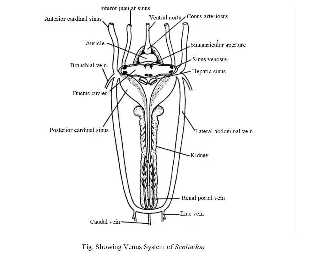
Blood returns to the heart from the anterior part of the body through the paired jugular and anterior cardinal sinuses. Blood from the posterior region is received through a pair of posterior cardinal sinuses. The anterior and posterior cardinal sinuses on each side combine to form a transverse sinus called the ductus cuvieri. The cardinal system can be divided into two parts, namely :
(1) Anterior cardinal system and
(2) Posterior cardinal system.
(1) Anterior Cardinal System
Blood from the head region (brain) returns to the heart through the veins of this system. It consists of a pair of internal jugular veins. Each internal jugular vein consists of the olfactory sinus, orbital sinus, post-orbital sinus, and anterior cardinal sinus.
Blood is transmitted through the anterior facial vein from the rostral region and enters the olfactory sinus. From there it goes to the orbital sinus. The orbital sinus is exposed to the anterior cardinal sinus through the post-orbital sinus. The anterior cardinal sinus enters the ductus cuvieri. The anterior cardinal sinus receives the hyoidian sinus and the 5 dorsal nutrient branchial sinus from the gills.
(2) Posterior Cardinal System
The caudal vein collects blood from the tail region and moves forward through the haemal canal. In the abdominal cavity, the caudal vein divides to form the right and left renal-portal vein, which divides into a sinusoid capillaries in the kidney. Along its entire length, the renal-portal vein acquires small parietal veins. Renal veins receive blood from the kidneys and then unite to form the posterior cardinal sinus. The two posterior cardinal sinuses open in the ductus cuvieri.
2. Hepatic Portal System
A significant number of small veins collect blood from the alimentary canal and its associated glands and later merge to form the hepatic portal vein. Lienogastric vein, anterior and posterior gastric veins merge with the hepatic portal vein.
In fact, the anterior and posterior gastric veins combine to form the hepatic portal vein. It is divided into capillaries in the liver. Blood is collected from the liver through another set of capillaries which later merged to form two large hepatic sinuses that are exposed to the sinus venosus.
3. Cutaneous System
Cutaneous system consists of a dorsal, a ventral and two pairs of lateral cutaneous veins. The inferior lateral cutaneous vein is connected to the lateral cutaneous vein near the anterior edge of the thoracic/pectoral fin. Each lateral cutaneous vein is usually combined with the branchial vein.
4. Ventral System
Ventral system consists of two sets of veins, namely :
(1) anterior ventral vein– which carries blood to the ductus cuvieri through the inferior jugular sinuses; and
(2) posterior cardinal vein– it supplies blood through the subclavian vein.
The veins of each inferior jugular are composed of the submental sinuses of the lower jaw, the hyoidean sinus and the ventral nutrient sinuses from the gills. The jugular veins of each inferior are exposed in the ductus cuvieri. The subclavian vein is also exposed in the ductus cuvieri on each side.
Two large lateral abdominal veins are formed with a small caudal vein and two iliac veins. The lateral abdominal vein is connected to the posterior part by a commissural vein. Anteriorly, the lateral abdominal veins merge with the branchial veins to form the subclavian vein which is exposed to the ductus cuvieri.
Arterial System of Teleosts
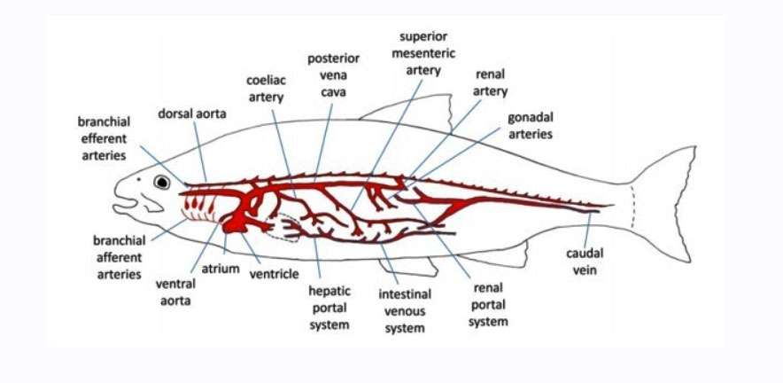
The ventral aorta moves forward and gives off four pairs of afferent branchial blood vessels of which third and fourth pairs originate from the same common location of the ventral aorta and supply blood to the third and fourth gills. These blood vessels travel to the holobranchs on each side and reach to the paired blood capilaries of the gill lamellae. In gills, blood is oxygenated and blood is collected through four pairs of efferent branchial arteries.
Blood circulation layout of bony fish
Each gill arch contains one efferent blood vessel, the first two of these originate dorsally from the gills and connects to form the first epibranchial blood vessel. The epibranchial arteries on both sides runs the posteriorly and join to form the dorsal aorta. The third and fourth efferent branchial blood vessels originate from the corresponding holobranch and join to form a short second epibranchial blood vessel which opens into the dorsal aorta.
A short common carotid artery originates from the first efferent branchial blood vessel, protrudes and divides somewhat to form an external carotid and an internal carotid artery. The carotid artery near its base receives blood from an efferent pseudobranchial artery coming from the pseudobranch. A cerebral artery is generated from the common carotid artery and supplies blood to the brain. The external carotid artery divides into numerous branches and supplies blood to the operculum, auditory region, and muscles of the jaw.
The internal carotid artery supplies blood to the snout and optic region. A small branch is formed from the internal carotid artery and goes along the midline towards the front and joins with the branch coming from the other side to form the circulas cephalicus. The dorsal aorta extends posteriorly below the vertebral column. The subclavian artery arises from the dorsal aorta just behind the second epibranchial artery and supplies blood to the pectoral fins.
The coeliaco-mesenteric artery arises from the dorsal aorta just behind the subclavian artery and progresses a little further, splitting into two branches called the coeliac and mesenteric arteries. The coeliac artery supplies blood to the anterior region of the intestine. On the other hand, mesenteric artery gives off branch and supplies blood to the liver, spleen, gonads, and to the rest of the alimentary canal.
The dorsal aorta reaches through the kidney and produces a few pairs of renal arteries on its lateral side. One of these pairs reaches into the two pelvic fins, and then the dorsal aorta continues posteriorly, becoming known as the caudal artery along the middle of the hemal canal, and gives off a few pairs of segmental arteries that expand into the muscle during its course.
The above description represents an ideal arrangement of arterial system in teleost. However, some variations in the arteries can be observed in different species of freshwater, such as the four pairs of afferent branchial arteries arise in the Catla catla that originate independently. However, in Mystus aor, Rita rita, Tor tor, Clarias batrachus, Heteropneustes fossilis, Wallago attu, Chitala chitala, the third and fourth arteries on each side originate from the same common place. In a very small number of species of fish, such as Rita rita and Heteropneustes fossilis, the second pair of afferent arteries originate in the same common way from the ventral aorta.
In some species, such as the Catla catla, Tor tor have a pseudobranch which arise from first efferent branchial artery and receives blood supply through the afferent pseudobranchial artery. Blood is collected through the afferent pseudobranchial artery that connects to the internal carotid artery. Mystus aor does not have a pseudobranch. In this case, the base of the internal carotid artery is swollen to form a labyrinth.
The alimentary canal and its associated glands receive blood supply from the branches of the coeliaco-mesenteric artery. The gonads receive blood from the coeliaco-mesenteric or the posterior mesenteric arteries.
Venous System of Teleost
Blood is collected from the head through the external and internal jugular veins which combine on each side to form the anterior vein. The internal jugular veins receive blood supply from the premaxillary, nasal, and eye regions. External jugular veins, on the other hand, collect blood from the maxillary and mandibular regions.
The anterior cardinal vein receives blood from the opercular and subclavian veins before opens into the ductus cuvieri. A single inferior jugular vein collects blood from the ventral surface of the pharynx and is exposed to the sinus venosus.
There is a single posterior cardinal vein in the teleost that reaches to the right kidney. The renal veins coming from both kidneys are exposed to the posterior cardinal vein and runs forward and is exposed into the sinus venosus.
The blood coming from the tail is collected through the caudal veins which gain some segmental veins and are exposed to the kidneys. Hepatic portal veins collect blood from different regions of the alimentary canal, spleen, swimbladder, and gonads and reach to the liver. Later, two hepatic veins are generated from the liver and supply blood to the sinus venous.
In Tor tor, this venous system represents the ideal venous system of teleost. However, some variations in the venous system can be observed in different species of freshwater fish. The inferior jugular veins are usually unpaired. But in some fish, such as Clarias batrachus, have two inferior jugular veins. In Tor tor, Catla catla, Wallago attu, the posterior cardinal veins are unpaired. However, in Clarias batrachus it is paired. Although the right posterior cardinal vein is more developed in this species.
Lymphatic System of Teleost Fishes
Like other vertebrates, fish collect lymph from all parts of the body through a system consisting of paired and unpaired ducts and sinuses, which ultimately return into the main bloodstream. The upper vertebrates have lymph nodes but are absent in fish.
The lymphatic system of lamprey and hagfish (Cyclostomata) is characterized by more numerous and more diffuse connection with the blood circulatory system than exists in other groups of fishes. Because of this close connection, the blood vessels are called the hemolymph system. The lamprey and hagfish have a large abdominal lymph sinus that enters into the lymphatic ducts of the kidneys and gonads.
There are several valve openings in the sinuses of the cardinal vein. The valves allow lymph flow to enter the veins and prevent a back flow of venous blood into lymph sinuses. The headcervical region of lamprey contains superficial and deep lymph sinuses where the valves of a lymphatic peribranchial sinus are connected to the jugular veins.
In the Elasmobranch, the lymphatic system contains much lymph vessels than the sinuses, but the cyclostomata and osteichthyes do not contain contractile lymph ‘heart’.
The sub-vertebral lymph trunk is situated in the hemal canal of the tail vertebrae which collects lymph fluids from the tail region. It then merge into the abdominal lymph duct which form a network of blood vessels with the lymphatic system.
The lymph-collecting vessels from the segmental musculature and intestinal organs flows into the sub-intestinal lymph trunk which in turn open into the cardinal sinuses near the site of origin of the subclavian artery from the aorta. Subvertebral lymph trunks extend into the head and where they collect lymph from the cranial and branchial regions.
The lymphatic system of fish is thought to be more likely to originate from the veins than the arterial part of the blood circulatory system. In Elasmobranchii, Chondrostei and Holostei, the complexity of its growth, development and number is gradually increased.
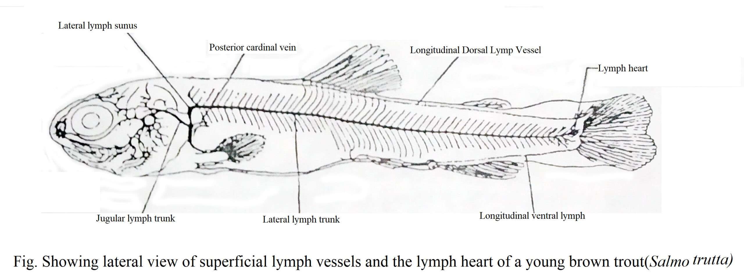
In teleostei, the arrangement of the lymphatic vessels is better than that of the terrestrial vertebrate, and the branches of the subcutaneous lymphatic ducts are more extensive. The lymph from the head region collects in the branched sinuses and flows into the sub-scapular sinuses of the pectoral region, where it is unite by fluid from the three main lymphatic ducts of the body-the dorsal, lateral, and vertebral subcutaneous lymph trunks.
Neural, arterial, and hemal sub-musculature lymph trunk collect the lymph fluids from body musculature. On the other hand, the lymphatic ducts of the visceral organs divide and form superficial and a deep systems. The deep visceral lymphatic duct absorbs fat from the intestinal mucosa and carries it to the ciliaco-mesenteric lymph trunk where the remaining lymphatic ducts are probably connected to the subvertebral trunk.
The lymph enters into the paired para-renal lymphatic duct from the bladder, gallbladder, abdominal part of the kidney, and other organs of the body cavities and subsequently ends in the pericardial sinus.
In Actinopterygii, the lymph reaches the main bloodstream through the anterior (cephalic) lymph sinus, which opens into the cardinal vein, and such conditions can be observed in conger eel(Comger) and freshwater eel (Anguilla).
The opening of the anterior lymph sinus connecting the blood and lymphatic system, also exists in the jugular vein, as seen in some fish Morays (Muraena), and in Pike (Esox), or in the posterior cardinal veins as in some members of the family Salmonidae (Salmo).
In conger eel (Conger) and freshwater eel (Anguilla), a false lymph heart is located in the cephalic sinus and keeps the lymph flow intact through the movement of the gills but a true lymph heart with valves and cardiac muscle contractile fibers occurs in the caudal region of both Anguilla and Salmo.
A small blister-like flattened structure appears in the hypural on the ventral of the last vertebra of the true lymph heart, which is covered by muscles and skin. They make contact with the lymphatic ducts and caudal veins of the body and are double-chambered and valved; they are thought to further venous flow.
Fish Blood as a Gas Carrier
Oxygen spreads from one liquid to another very slowly. Red blood cells have appeared in fish and other vertebrates to achieve high efficiency in gas transport. This is why one volume of blood can carry 15-25 times more oxygen than water. 99% red blood cells and 1% plasma contribute to this oxygen transport. The red blood cells of fish and other vertebrates contain a type of pigment called hemoglobin. In its presence, the blood turns red and acquires the ability to transport oxygen. In most vertebrates, the molecular weight of a hemoglobin is about 65,000. The oxygen carrying capacity of hemoglobin of some fish is given in the table below:
Table :The oxygen carrying capacity of hemoglobin of some fish
|
Species |
Oxygen Carrying Capacity (Percentage of Blood Volume) |
Number of red blood cells (per cubic mm) |
Iron content (mg per 100 cc) |
|---|---|---|---|
|
Goosefish(Lophius (Sedentary) |
5 |
867,00 |
13.4 |
|
Mackerel-(Scomber) (active) |
16 |
3,000,000 |
37.1 |
The table above shows that the maximum oxygen holding capacity of Mackerel is 18% but that of mammals is 20%. Oxygen is taken up, transported and released by red blood cells. So we can call this process carriers, loading and unloading.
Table: Differences between plasma and serum
|
Plasma |
Serum |
|---|---|
|
Plasma is the clear, yellowish portion of blood, lymph, or inter-muscular fluid that holds blood cells. |
Serum is the clear, yellowish liquid that is obtained after all the solid and liquid components have been separated from it after blood clotting. |
|
It contains fibrin and prothrombin proteins and other soluble blood clotting components. |
It does not contain proteins called fibrin and prothrombin. |
|
It helps in blood clotting. |
It does not help in blood clotting. |
Table-6.3: Differences between different types of fish blood cells
|
Red blood cells or Erythrocytes |
White Blood Cells or Leukocytes |
Platelets or Thrombocytes |
|---|---|---|
|
Most fish have red blood cells with round or rectangular nuclei that are located in the center of the cell. |
Their nuclei are round, oval or different in shape. |
It is round, oval or spindled shaped. |
|
It is yellowish red in color. |
It is white in color. |
It is white in color. |
|
In blood of fish, the number of red blood cells per cubic millimeter is 20,000-30,000,000. |
The number of White Blood Cells per cubic millimeter is 20,000-100,000. |
The number of platelets per cubic millimeter blood of fish is half of leukocytes. |
|
They carry oxygen and play an important role in respiration. |
They help prevent disease. |
They help in blood clotting. |
|
It is produced from hemoblasts. |
It is also produced from hemoblasts. |
It originates from the mesonephric kidney of fish. |

