Scoliodon is an Indo-Pacific species which belongs to the family Ceacharrhinidae under order Carcharhiniformes of Class Elasmmobranchii. It has an elongated and spindle-shaped body which tappers at the end. The head is dorso-ventrally compressed with laterally compressed trunk and tail. The mouth is situated on the ventral side and the entire body is covered by placoid scales.
Systematic Position
- Phylum: Chordata
- Subphylum: Vertebrata
- Class: Elasmobranchii
- Order: Carcharhiniformes
- Family: Carcharhinidae
- Genus:Scoliodon
The circulatory system of cartilaginous fish such as Scoliodon is composed of blood, heart, arterial system and the venous system. In Scoliodon, there are two distinct arteries in the arterial system, namely-
1. Afferent branchial arteries; and
2. Efferent branchial arteries.
The arterial system of Scoliodon is briefly described below:
1. Afferent Branchial Arteries
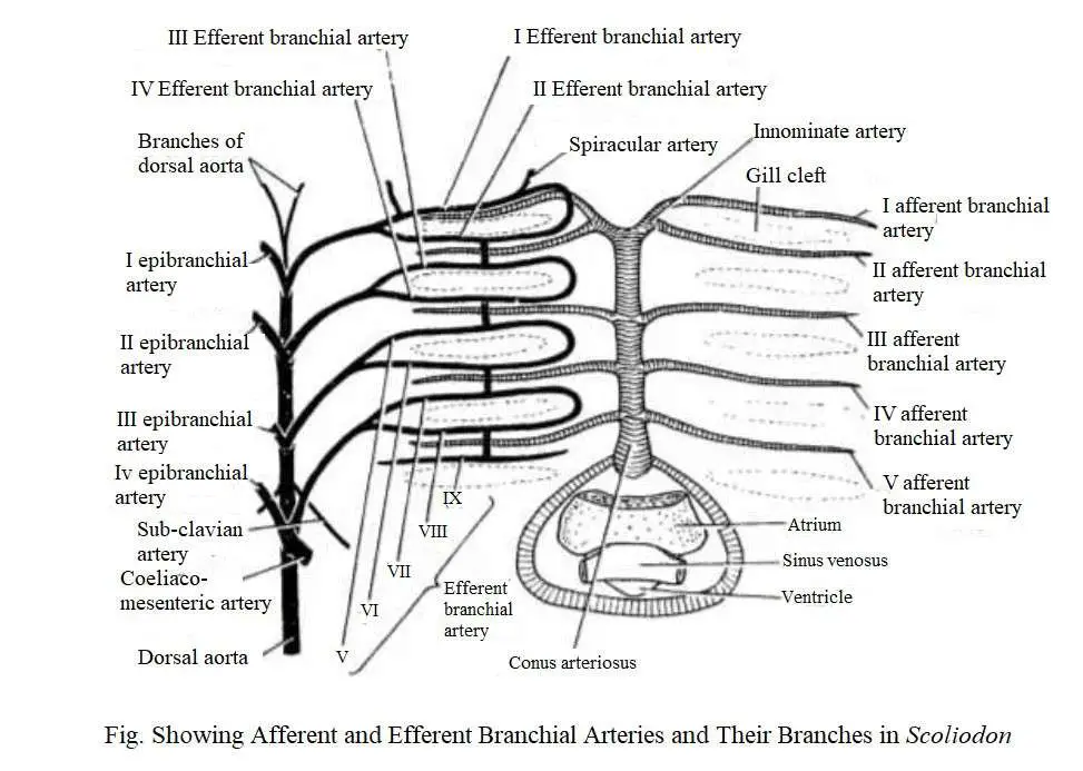
Afferent Branchial Arteries begin from the ventral aorta and carries oxygen-free blood to the gills for oxygenation. The ventral aorta is situated on the ventral surface of the pharynx. It extends up to the posterior boundary or hyoid arch.
The ventral aorta is divided into two branches called the innominate arteries, each of which re-divides into two branches to form the 1st and 2nd afferent branchial arteries. The 3rd, 4th and 5th afferent branchial arteries originate from the ventral aorta.
Each afferent artery originates from a ventral aorta through an independent opening except for the 1st and 2nd afferent branchial arteries which are exposed to the same common opening.
2. Efferent Branchial Arteries
Efferent Branchial Arteries arise from the gills and carries oxygenated blood to different parts of the body. The efferent branchial artery divides into capillary blood vessels in the gills. Blood is collected from the gills by efferent branchial arteries.
In Scoliodon, there are 9 pairs of efferent bronchial arteries that are evenly distributed on each side. The first 8 arteries form a series of four complete loops around the first four gill slits.
The 9th efferent branchial artery collects blood from the hemibranch of the 5th gill pouch and from where the blood is poured into the 4th loop. In addition, the shorter longitudinal connector connects the four loops. These are re-connected with each other by a network of longitudinal commissural blood vessels called the lateral hypobranchial chain.
An epibranchial artery originates from each efferent branchial loop. The four pairs of epibranchial arteries join along the mid-dorsal line to form the dorsal aorta. The 9th efferent branchial artery has no epibranchial branch. However, it joins with the 8th efferent branchial artery.
Anterior Arteries
The head region receives blood supply from the 1st efferent branchial artery and partly from the proximal end of the dorsal aorta. The following arteries originate from the 1st efferent branchial artery (hyoidian efferent), viz. (a) external carotid (b) afferent spiracular (c) hyoidean epibranchial which gets blood from a branch of dorsal aorta.
The external artery receives blood from the first collecting loop and subsequently divides to produce a ventral mandibular artery and a superficial hyoid artery.
The ventral mandibular artery produces branches to the muscles of the lower jaw and the superficial hyoid artery which supplies blood to the 2nd ventral contractile muscle, the skin and subcutaneous tissue below the hyoid arch.
The afferent spiracular artery originates from the medial space of the hyoidian efferent artery and enters the cranial cavity as it progresses forward as the spiracular epibranchial artery. Just before its entry to the cranial cavity it sends large ophthalmic arteries to the eye ball.
As the spiracular epibranchial artery enters the cranial cavity, it connects to a branch of the internal carotid to form the cerebral artery. It later divides to form an anterior and a posterior cerebral arteries, which supply blood to the brain.
The hyoidian epibranchial artery runs forward and enters the posterior boundary of the eyeball, and it acquires an anterior branch from the dorsal aorta. It later splits to produce:
(1) the stapedial artery, which re-divides to form the inferior orbital artery and the superior orbital artery. The superior orbital artery moves forward and enters the superficial tissue above the 6 eye muscles and the auditory capsule.
From the superior orbital artery a large buccal artery arises and progresses as the maxillo-nasal artery. A few branches originate from the maxillo-nasal artery and enter the muscles of the upper jaw, the olfactory sac and the rostrum.
(2) The internal carotid artery passes inwards and enters the cranium where divides into two branches. One of the branch unites with its fellow from the opposite side and the other branch unites with the stapedial.
Dorsal Aorta and its Branches
The epibranchial arteries converge to form the dorsal aorta and move posteriorly. It is situated on the ventral side of the vertebral column. It extends up to the tip of the tail as a caudal artery. The dorsal aorta along the antero-posterior direction produces the following arteries, viz.
(1) Several buccal and vertebral arteries, which originate from anteriorly;
(2) Subclavian arteries-originate from the fourth epibranchial artery. An epicoracoid artery originates from the subclavian artery. The subclavian artery subsequently re-divides into three branches, namely-
(i) the branchial artery which enters the pectoral girdle and pectoral fins;
(ii) an antero-lateral artery which enters the body musculature;
(iii) a dorso-lateral artery which enters the dorsal musculature;
(3) A large ciliaco-mesenteric artery arises from some posterior part of the origin of the 4th epibranchial artery. It is further divided into two parts, such as a smaller coeliac artery and a larger anterior mesenteric artery;
(4) Lienogastric artery-it originates from the posterior part of the ciliaco-mesenteric artery and divides into the following branches, viz.,
(I) an ovarian (in female) or spermatic artery (in male) that enters the genital organs;
(ii) a posterior intestinal artery– which enters the posterior part of the intestine;
(iii) a posterior gastric artery-which enters the posterior part of the cardiac stomach;
(iv) a splenic artery-which enters the spleen;
(5) Paired parietal arteries – which originate from the posterior part of the subclavian artery. Each parietal artery is divided into a dorsal and a ventral parietal artery.
The dorsal parietal artery supplies blood to the dorso-lateral musculature, the vertebral column, spinal cord, and the dorsal fin. The arterial parietal artery supplies blood to the ventral muscles and the peritoneum. From this paired parietal artery, the renal artery enters the kidney.
(6) A pair of iliac arteries that extend to the pelvic fin and become known as the femoral arteries.
Hypobranchial Chain
A network of slender arteries arising from the loop of the ventral ends of the efferent branchial artery forms a lateral hypobranchial chain. From it, four commissural blood vessels are formed and join the ventral wall of the ventral aorta to form a pair of median hypobranchials which are connected to each other by transverse blood vessels.
Posteriorly, the median hypobranchials unite to forms a median coracoid artery from which the coronary artery and a pericardial artery originate. The common epicoracoid artery originates from the pericardial artery and later divides into the right and left epicoracid arteries, each of which is connected to a subclavian artery.

