The heart in all animals is a muscular organ. In human, it is placed in the position of the thoracic cavity just behind the sternum and is slightly tilted to the left. Thus, the heart-beat is felt on the left side. It pumps blood throughout the blood vessels to various parts of the body by repeated rhythmic contraction.
The heart is a reddish-brown muscular organ which is somewhat cone-shaped with the broader upwards end called the base and the blunt end called the apex lying on the diaphragm.
The weight of the heart in adult male and female is about 280-340 grams and 280 grams, respectively. In adult, it measures about 12 cm in length and 9 cm in breadth.
The heart is surrounded by a double-walled structure called pericardium. It consists of an outer fibrous and an inner serous pericardial membranes, also known as visceral layer.
The inner layer is tightly affixed to the wall of the heart. The outer layer is composed of relatively inelastic connective tissue, and is also called parietal layer. It prevents distension of the heart.
The space between the pericardial membranes is filled with a fluid, known as pericardial fluid which acts as cushion. This fluid protects the heart against shocks and also minimizes the friction on the outer surface of the beating heart.
Hear Chambers
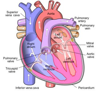
The heart of man are made of four chambers, of which, two upper chambers are called the atria or the auricles, left auricle and right auricle and two lower chambers are called the ventricles, left ventricle and right ventricle.
Atria or Auricles
Two auricles are separated by intra-atrial septum whereas the two ventricles are separated by intra-ventricular septum. The auricles and ventricles are completely separated from each other by a fibrous structure, known as fibro-tendinous ring.
Right auricle receives de-oxygenated blood from the body by superior and inferior vena cava. This chamber also gets more carbon dioxide-rich blood from heart itself by coronary sinus. The left auricle or atrium gets oxygenated blood from the lungs by four pulmonary veins ( 2 from each lung).
Ventricles
Ventricles are thick-walled structures separated from each other by the interventricular septum. The left ventricle is more narrowed and longer than the right ventricle. It forms the apex of the heart. The ventricles have more wide and muscular walls than the auricles. In this case, the walls of the left ventricles are at least three times thicker than those of the right.
From right ventricle, pulmonary artery arises. It carries venous (Carbon dioxide-rich blood) blood from heart to lungs. Aorta arises from the left ventricle which carries oxygenated blood from heart to all parts of the body.
The ventricle pumps the blood to lungs and body parts. The left ventricle has a circular opening of the systemic aorta guarded by three aortic or semi lunar valves. The right ventricle has a rounded opening of pulmonary artery guarded by three pulmonary or semi lunar valves.
The heart wall contains the following three layers:
- An outer epithelium lining called epicardium;
- A middle muscular layer called myocardium and
- An inner endothelium lining layer, known as endocardium.
The epicardium is made up of soft connective tissue and its outer layer is thin and transparent while the myocardium is composed of cardiac muscle tissue. The majority of the cardiac wall contains cardiac muscle tissue. These tissues help in pumping action of the blood.
The thickness of the myocardium varies in different parts of the heart. The atrial myocardium is thin as it has to do small amount of work in pumping blood to ventricles only. But the ventricular myocardium is thick as the ventricles do most of the works of the heart for pumping the blood throughout the body by way of the blood vessels. The ventricular myocardium sends out projections in the ventricular.
The endocardium has thin layer which overlaps the connective tissue layer. It gives a soft lining for the heart chambers and envelops the valves of the heart.

Diagram of the Human Heart: image credit: wikipedia.org
Valves of the Heart
There are different openings or orifices are present in the heart. These openings are guarded by valves. To circulate the blood strictly in one direction and to prevent any admixture between arterial and venous blood, four sets of valves are present in the heart.
Each atrium opens into the ventricle of its own side through atrio-ventricular apertures. The right aperture is guarded by the tricuspid valve and the left by bicuspid valve or mitral valve. Part of each valve is attached to the fibro-tendinous ring which surrounds the two atrio-ventricular apertures. The free margins are attached to the papillary muscles of the ventricles by strong chordac tendineae. The base of the aorta and pulmonary artery are guarded by aortic or semilunar valves.
The heart is made up of involuntary striated muscle which is known as cardiac muscle. Cardiac muscles share some features with the muscles namely the smooth type and the skeletal type but have some features that make difference to others. The features are:
- Cardiac muscles functions are controlled involuntarily.
- The cells of the muscle branch randomly and twined with each other, thus resembling a three dimensional mesh.
- The cardiac muscle cell contains many parietal arranged myofibrils.
- The cardiac muscle contains branched cells which are connected by intercalated discs.
- The protective wall of the heart contains cardiac muscles. In this case, they are concentrated in the myocardium part.
- The major function of the cardiac muscle is pumping blood to the blood vessels for circulation throughout the body.
Some special type of tissues is present in the heart. They are known as junctional tissues of the heart. It is mainly of four types:
1. Sino-atrial nodes (S.A. node): It is present at the base of the superior vena cava in the right atrium.
2. Atrio-ventricular node (A.V. node): It is present in the right atrium just above the atrio-ventricular septum.
3. Bundle of His: These are a bundle of fibres originating from A.V. node which passes along the intraventricular septum.
4. Purkinje fibres: These are fibrous structures that arise from the branches of bundle of His and end within the ventricular muscle. The function of these junctional tissue is to initiate and to conduct cardiac impulse. S.A. node initiates 70-80 times cardiac impulse which regulates the rhythmicity of the heart beats 70-80 time (average 72 times) per minute. Thus S.A. node is known as pace maker of the heart.
Blood Circulation Through the Heart
The heart is the central receiving and pumping organ of the circulatory system. The heart pumps around 2000 gallons of blood everyday and beats nearly 100000 times per day. Due to its constant rhythmic contraction-relaxation blood can flow throughout the body. The contraction and relaxation of the heart termed as systole and diastole respectively.
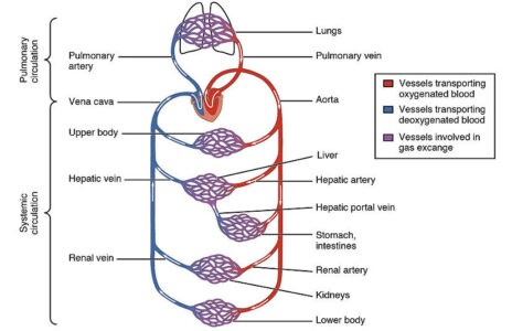
All parts of the heart do not contract or relax at a time. When atria are contracted then the ventricles become relaxed and this happen vice-versa. As a result, continuous flow maintain throughout the heart. The human heart has two routes through which blood pass, in and out of the heart. This type of circulation is called the double circuit circulation. During blood circulation the right and left sides of the heart work together.

Image Showing Blood Circulation: Image credit: wikimedia commons
Blood Circulation Through Right Side of the Heart
- Blood enters the heart through two large veins, the superior and the inferior vena cava discharging O2-poor blood from the body into the right atrium of the heart.
- As the chamber shrinks, blood streams from the right atrium into the right ventricles through the open tricuspid valve.
- The tricuspid valve is closed after when the ventricle is full. This helps to keep blood from streaming in reverse into the atria while the ventricle contracts.
- As the ventricle shrinks blood goes away the heart through the pulmonic valve into the pulmonary artery and to the lungs where it is oxygenated.
Blood Circulation Through Left Side of the Heart
- The aspiratory vein discharges O2-rich blood from the lungs into the left chamber or atrium of the heart.
- As the chamber contracts, blood streams from the left chamber or atrium into the left ventricle through the open mitral valve.
- The mitral valve closes while the ventricle is full. This keeps blood from streaming in reverse into the atrium while the ventricle contracts.
- As the ventricle contracts, blood goes away the heart through the aortic valve into the systemic aorta and to the body.
