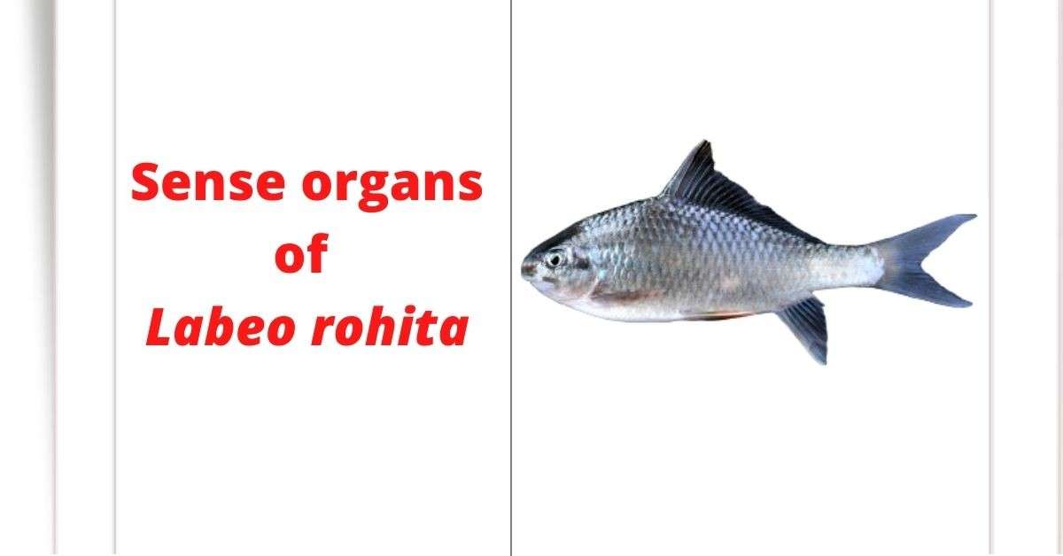
The sense organs of Rohu fish (Labeo rohita) consist of eye, internal ear, lateral line sensory organ and olfactory organ. These organs are made according to the general design of bony fish.
Eye
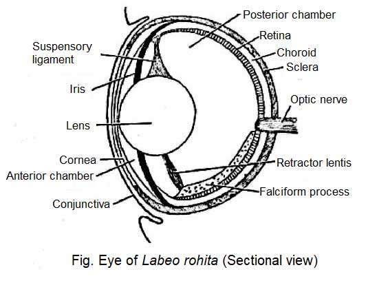
The basic structure of the eyes of Rohu fish (Labeo rohita) is similar to the eyes of other vertebrates. The eyeball is located in the orbit of the eye. The layers of the eyeball are three: the outer layer is called the sclera, the middle layer is called the choroid and the inner layer is called the retina.
Sclera protects the eyeball because it is rich in cartilage. Choroid is rich in blood vessels and glandular and retina is rich in sensory cells. The visible part of the sclera at the tip of the eyeball is transformed into a transparent and thin screen to form the cornea. The cornea is covered with a transparent covering called the conjunctiva. Like other vertebrates, the part of the choroid layer behind the cornea forms the pigmented iris.
The central opening in the Iris is called the pupil. A round lens hangs from the wall of the eyeball to the inside of the eyeball with the help of a suspensory ligament. The small space of the eyeball between the lens and the cornea is called the anterior chamber. A watery fluid is present in this anterior chamber, known as aqueous humor.
The large space behind the lens is called the posterior chamber. This chamber is filled with a jelly-like liquid called vitreous humor. A fold through the choroid layer near the entrance to the optic nerve in the eyeball extends to the posterior edge of the lens. This is called falciform process. This organ is attached to the lens by the retractor lentis muscle. It is a kind of contractile muscle. In the contraction of this muscle, the lens is closer to the retina. The position of the lens is controlled by this muscle.
The retina of the Rohu fish is flat and the lens is round. The front of the eye is very small because the cornea is located close to the body of the lens. Therefore, under normal conditions, they become myopic. But to see distant objects requires adjustment/adaptation. Retractor lentis play an important role in fulfilling the prerequisite of adaptation. Retractor lentis is a type of contractile muscle that attaches the lens to the falciparum process.
Inner Ear
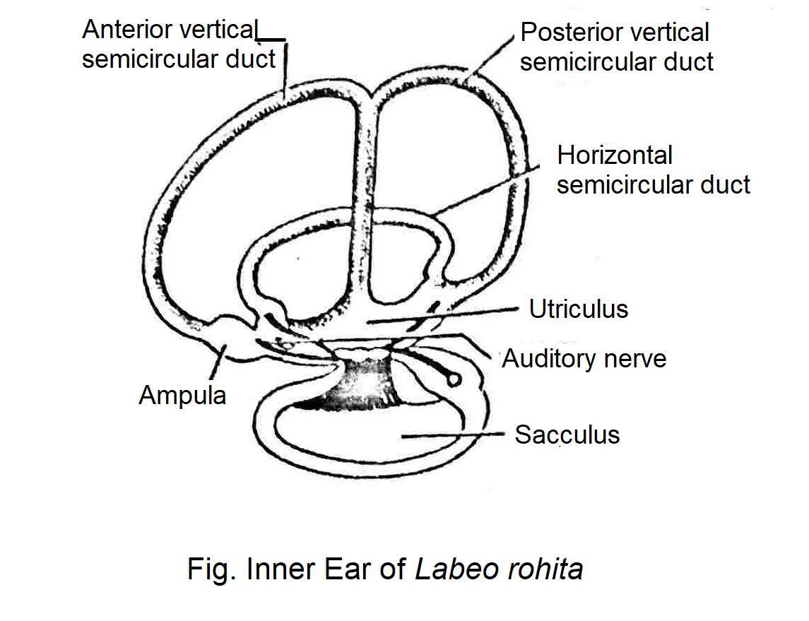
Fish do not have external ear or middle ear. Their inner ears are similar to those of other vertebrates. The inner ear consists of a sac-like membranous labyrinth. It has three semicircular ducts and three chambers. These three chambers are Utriculus, Sacculus and Lagena. Each chamber contains an otolith. The otoliths are located in a fluid called endolymph in the inner ear.
There are three types of autoliths. These include Lapillus, Sagitta and astericus. Among them, lapillus is found in Uticulus, Sagitta in Saculus, and Astericus in Lagina. Sajita is the largest of them in size. The semicircular duct is connected to the utriculus. One end of each semicircular tube swells to form an ampulla. The brain senses when the sensory hair existing in the cells comes in contact with the autolith. The auditory nerve connects the brain to the inner ear. In addition to hearing, the inner ear helps maintain the balance of the body.
Lateral Line sensory Organ
From the back of the brain there are two lateral lines on either side along the length of the body. This line connects with the narrow ducts in the body through numerous pores. Numerous sensory cells or neuromast are located inside the narrow ducts. Each cell is connected to a nerve. The lateral sensory cells communicate with the lateral branch of the tenth cranial nerves as a whole.
Olfactory Organ
The nasal sacs form the olfactory organ. The olfactory organ is not directly connected to the oral cavity, but each nasal sac communicates with the outside through the external nostrils. The olfactory cells of the nasal sac are capable of perceiving the odor of various objects as chemoreceptors and olfactory cells.


