Respiration is a biochemical process by which food in the cell is oxidized with the help of oxygen to produce energy and carbon dioxide (CO2) is released as a by-product. All kinds of biological function require such energy. Carbohydrates are mainly involved in energy production. In the absence of carbohydrates, this energy is produced by oxidizing fats and proteins. This reaction is shown below through chemical equations.
Respiration is a feature of life and an indicator of all biochemical activities in the body. Respiration involves the exchange of two gases, oxygen (O2) and carbon dioxide (CO2). The respiratory system is made up of all the organs that are involved in the function. Based on the presence of oxygen, respiration is divided into aerobic and anaerobic.
In aerobic respiration, free oxygen is taken from the environment and carbon dioxide is emitted as energy is produced. This type of respiration occurs in most plants and animals. Organisms that breathe this way are called aerobes. Lactic acid is produced when glucose is metabolized in anaerobic respiration. In this case, no oxygen is required and no carbon dioxide is formed. Such respiration occurs in certain bacteria, parasites, etc. Such organisms are called anaerobes. The absence of oxygen in an organism is called anaerobiasis.
Like other animals, adequate oxygen supply is required to fish tissues for oxidataion and energy production. A respiratory organ has formed in animals to receive oxygen for intracellular oxidation and to sustain life and to emit carbon dioxide.
Oxygen and carbon dioxide are exchanged between blood and water (air) through the respiratory organs. This type of breathing is called exhalation. Through the exchange of blood and body tissues and gases, energy is generated in the cell which is called inhalation.
In most animals, the presence of special organs to control respiration can be observed. The respiratory system is made up of these organs. There are four main types of respiratory organs in the animal kingdom. These are:
- Integument;
- Gills;
- Trachea and
- Lungs.
Fish have well-developed respiratory organs. The physiological processes of respiration of different fish are roughly similar to those of the upper vertebrae. However, there are differences in the respiratory organs. Fish are the main aquatic vertebrates and take up most of the dissolved oxygen from the water. Some fish have the ability to breathe air. The gills are the main respiratory organs of fish. However, some bony fish have respiratory organs that help them breathe air. In air breathing fish, gills play a complementary role. As an efficient respiratory organ, the gill can use up to 70% of the dissolved oxygen in the water flowing through it. In humans, 25% of the oxygen in the lungs can enter the pulmonary cavity. The ability of the gills as a respiratory organ in fish depends on two factors, viz
1. The nature of the gill structure and capillary blood vessels;
2. Immerse the gills in a running stream of water so that fresh oxygen can always come in contact with the gills.
Location of Gills in Fishes
A row of vertical opening in the side wall of the pharynx indicates the presence of gills. Each pharyngeal opening enters a flattened gill pouch that connects to the outside through the gill openings. The gill pouches are separated by interbranchial septum. These septum or interstices carry gill filaments to its opposite wall. At the tip of the head, between the eyes and the thoracic fins, there is an external gill openings.
The gill openings are usually small, but in the Cetorhinus (huge basking shark), the openings are very large, extending from the top to the bottom of the body. Other gill openings are located in the posterior branchial arches. Most cartilaginous fish have five gill openings on each side in addition to the spiracle, but some sharks, such as the Hexanchus, have six gill openings except the spiracle, and the Heptranchias have seven pairs of gill openings.
In most cartilaginous fish, especially sharks, the gill pouch is exposed to the outside through individual external gill openings, and the gill openings are covered by posterior dermal folds.
Bony fish have interpharyngeal gill openings but they are not individually exposed to the outside. All of these openings are exposed to a common branchial chamber that is covered by a movable operculum. Each branchial chamber is exposed outside by a large opening. Opercula of the two sides overlap or merge. In some Actinopterigians, the rate of convergence of this overlaping is so high that the opercular opening is reduced to a small bilateral round opening (e.g. eel fish).
The operculum consists of four broad and flattened bones that may or may not have long branchiostegal rays on the ventral side. Among cartilaginous fish, operculum is present in Holocephali. This structure of oparculam in this fish is the interstitial condition of cartilaginous and bony fish. In this case, the actual interbranchial septum is smaller and the gills are in a normal branchial chamber. On the outside of this chamber, like the operculum of bony fish, it is covered by a skin-like fringe. Each branchial chamber is exposed outside through an opening. The bony fish has four gills on each side of its head. These gills are covered by operculum or gill covers which are exposed on the outside by an opening.
Types of Gills
There are two types of fish gills based on location, viz
(1) Internal gills– In this case, the gills are placed inside the gill pouch (shark) or inside the branchial chambers (holocephalan and bony fish).
(2) External gills – External gills develop in many fish for respiration in the larval stage. On the basis of development, it is again divided into two types, viz
(a) True external gills – They do not depend on the internal gills but develop as a result of development of skin. In the larval stage, Polypterus and Lepidosiren have true external gills. The number here is four pairs. This external gill becomes extinct as it matures, but at the the adult stage, Protopterus also contains external gill remains. In Polypterus, leaf-like external gills are present located on the gill opening.
(b) Prolongation from the internal gills: This type of gill is formed by elongating the inner gill-filament and it is located outside the body. Such gills are found in some Selachian and embryos of some oviporous bony fish. Long fibrous external gills are made up of from the wall of the gill opening of embryos in cartilaginous fish. Sea water flows through such type of fibrous structures and plays a role in respiration.
In the viviparous cartilaginous fish (Selachian), these organs also participate in the absorption of nutrients. In the larval stage of some oviparous bony fish (Gymnarchus, Clupisdis) and other fish it plays a role in respiration. In the Gymnarchus, a thin fibrous growth is formed from the opercular end of the internal gills and acts as an external gills.
The gill is divided into four types based on its structure and function, viz
- Hemibranch,
- Pseudobranch,
- Holobranch and
- Lophobranch.
(1) Hemibranch: The gill pouches are separated by the interbranchial septum. The front and back walls of each septum carry rows of gill filaments. The row of gill filaments on one side of the interbranchial septum is called the hemibranch. Hemibranch is divided into two types, viz
(a) Mandibular Hemibranch: The mandibular hemibrunch usually helps to close the spiracle and it receives oxygenated blood. The mandibular hemibranch regulates blood pressure in the ophthalmic arteries by increasing the oxygen concentration in the blood of the brain and acts as an endocrine gland. Most of it is made up of acidophilic cells. In the Lepisosteus, the mandibular pseudobranch and the hyoidian hemibranch are very close together and it receives blood from the afferent hyoidian artery and the 1st gill arch efferent artery. In sturgeon (Acipenser), there is no contact between mandibular pseudobranch and hyoidian hemibranch. In Amia, there is no direct contact between the pseudobranch and the effernet or afferent blood vessels. In this case, the pseudobranchial blood vessel connects the orbital and ophthalmic blood vessels. Polypterus does not have mandibular pseudobranch.
(b) Hyoidean Hemibranch: Most fish have such gills. Most gills of Elasmobranchii are of the hemibranch type. In shark, each gill arch holds one afferent and two efferent blood vessels. In this case, 5-7 pairs of gill pouches are arranged on each side of the head and in Rays (Rajifornes) it is arranged in a linear direction. Sharks and Rays have an extra opening, called a spiracle. It has no role in respiration. In Selachian, it is extinct or bud like. In holocephalan, it is adjacent to the operculum. Most Actinopterygii do not have it. Acipenser, Lepisosteus, Polyodon and Amia have such gills. In Scaphirhinchus, it is greatly reduced. Polyopterus has mandibular pseudobrach and hyoidian hemibranch. Other crossopterygian (except Lepidosiren) have hyoidian hemibranch. In Latimaria it is small.
(2) Pseudobranch: When the hemibranch loses its actual respiratory capacity, it is called a pseudobranch. Many Actinoterigians, such as the Catla catla, have a hyoidian pseudobranch with a single gill filament in front of the first gill.
The pseudobranch is covered by free or single-layered mucous membranes. It develops entirely in the early embryonic stage and in the embryonic stage, it plays a role in respiratory function but in adulthood, it has no role in respiration. It receives oxygenated blood directly from the dorsal aorta and it has contact with the intra-carotid artery and blood vessels. It increases the concentration of oxygen in the blood and travels through the intra-carotid arteries to the brain and eyes, such as Wallago attu, Mystus aor, Notopterus notopterus, Channa.
In trout, it has a comb-like structure. In the case of perch, it is covered by a highly depleted pharyngeal epithelium. In Cod fish, pseudobranch are completely covered by the pharyngeal epithelium and form a glandular organ called the rete mirabile. Although it originates from the depths of the pharyngeal tissue, it also originates from the fine formation of glandular tissue. In the case of Amia, it is reduced and covered by the pharyngeal mucous epithelium. Pseudobranch plays an important role in filling the swimbladder and controlling intraocular pressure.
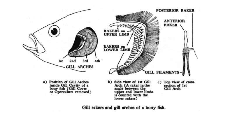
(3) Holobranch: When an entire gill consists of two hemibranch, it is called a holobranch. The holobranch contains an interbranchial septum (reduced in advanced fish) of which the front and back wall contain gill filaments with rich capilary blood vessels. An entire gill or a holobranch consists of cartilage or bony gill arch. Each arch has gill rakers on the inside and plate-shaped filaments with rich in blood vessels on the outside.
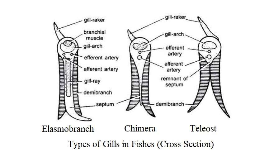
Elasmobranch usually have 5 pairs of gill openings but bony fish have 4 pairs of gill openings and there are no spiracles. The teleost develops a single external branchial opening, forming an operculum covering the gills on each side of the head. These fish have a decrease in the interbranchial septum and the conjunctival efferent branchial artery of the elasmobranch has become a single efferent blood vessel.
(4) Lophobranch: Sea horse (Hippocampus) and pipe fish (Syngnathus) have reduced gill filament which form a rosette-like hair follicle structure. These clusters are associated with small reduced gill arches. Such gills are called cluster gills or lophobobranch.
Gills of Different Fishes
The number, location and function of gills vary in different fishes. The following are the descriptions of gills in the major fish groups:
Gills of Lamprey (Petromyzonidae)
There are 6 pairs of gills on each side of the lamprey. These gill pouches are released into the pharyngeal cavity. Each pouch is divided by a diaphragm adjacent to the body wall. In addition to the diaphragm on the inner edge, the gill pouch has radially arranged gill filament which has small folds from one end to the other, increasing the respiratory level. The gill pouches have a cartilaginous structure, called branchial baskets. Through this, communication with the outside is achieved through the gill opening in the gill chamber.
Lamprey lives as parasite most of the time, so they do not use sucking mouths to breathe because they are attached to host fish or other objects such as rocks. In the lamprey, water enters the respiratory tract through the gill pouch and exits in the same way. In marine lampreys, the gill pouch is seen to contract and expand 50-70 times per minute while it is attached to the prey, but in river lampreys (Lampetra fluviatilis) it is seen to be 120 to 200 times per minute.
Respiratory water enters the gill pouch through the flow of water and the water is expelled by the contraction of the branchial compressor muscle adjacent to the branchial basket and the division of the inside of the pouch and the contraction of the sphincter around each gill opening. Water also enters through the contraction and dilation of the nasopharyngeal sac.
Gills in Hagfish (Myxiniformes)
In this case, the gills are transformed into gill pouch. The pouch of gill region are exposed in the pharynx. They have 6-15 pairs of gills. Myxine contains 6 pairs of gill pouch. These pouches are not exposed to the outside through an opening like lamprey, and each pouch forms a long emission tube. The 6 emission tube on each side then join together to form a common tube and are exposed to the outside through a single gill opening.
Gills in Cartilaginous Fish
In the gill pouch, there is a respiratory organ called gills. In different sharks, structure of gill pouches are of different types. They have 5 pairs of gill pouch on the lateral side of their head. Each gill pouch maintains communication with the pharynx through single intra-branchial openings and 5 external gill openings.
A row of horizontal branchial lamellae is produced from the lining of the mucous membrane of the gill pouch. These lamellae are enriched with excessive amounts of blood vessels. At the front or each gill pouch, there is a set of branchial lamellae and at the back, another set at of branchial lamellae. The gill pouches are separated by the interbranchial septum. The interbranchial septum tends to be longer in length than the branchial or gill lamellae. Each interbranchial septum has one visceral arches at the pharyngeal end.
The back of each arch carries the anterior lamellae of gill pouch and the rear set lamellae of the next gill pouch. The first gill pouch is between the hyoid and the 1st bronchial arch and the 2nd gill is located between the 1st and 2nd branchial arches, the 3rd gill pouch is located between the 2nd and 3rd branchial arches, the 4th gill pouch is located between the 3rd and 4th bronchial arches and 5th gill pouch is between 4th and 5th branchial arches. Two types of gills can be seen in sharks- Namely:
- Holobranh
- Hemibranch
1. Holobranch: This type of gill has 2 sets of gills or gill lamellae. That is why they are called complete gills or holobranch. The first 4 branchial arches carry holobranch.
2. Demibranch or hemibranch: This type of gill has a set of gills or gill lamellae. The hyoid arch carries a single hemibranch. The last branchial arch has no gills. During the respiration, the floor of the buccal cavity goes down or down and the mouth is exposed. Then water enters at a rapid rate and fills the overly stretched buccal cavity. Immediately after this, the mouth and pharynx become constricted. As a result, the water then enters the gill pouch and after exchanging the gas, the gill comes out through the gill opening. When the mouth is engaged in other work (catching prey), the spiracle provides a helpful pathway to enter water for the respiration.
Gills in Bony Fish
In this case, there are four pairs of gills on the back of the head, four on each side. The gills on each side are covered by gill covers or oparculam. The following is a description of the structure of gills:
The main respiratory organ or the ideal structure of the gills
The gills are the main respiratory organs of fish. There is a row of crevice-like openings in the side wall of the pharynx, the first of which is called a spiracle. It is located between the mandibular and the hyoid arch. The second or hyoidean cleft is located between the hyoid arch and the first branchial arch. The remaining gill openings are located between the posterior branchial arches. From the anterior and posterior walls of each gill opening, a blood-rich, fibrous outgrowth is produced where dissolved oxygen and carbon dioxide are exchanged. In addition to gills, skin and swimbladder act as respiratory organs.
Each gill structure looks like a comb and its gill arch has rows of gill filaments. Each gill arch carries two rows of gill filaments. The surface of each gill filament has numerous small folds that increase the overall surface area of the gill for gas exchange. The respiratory area of the fish gills depends on the size and number of gill filaments. Respiratory areas are developed based on fish habits. In fish, the principle of respiration of gill is almost the same.
Structure of a Teleostean Gill
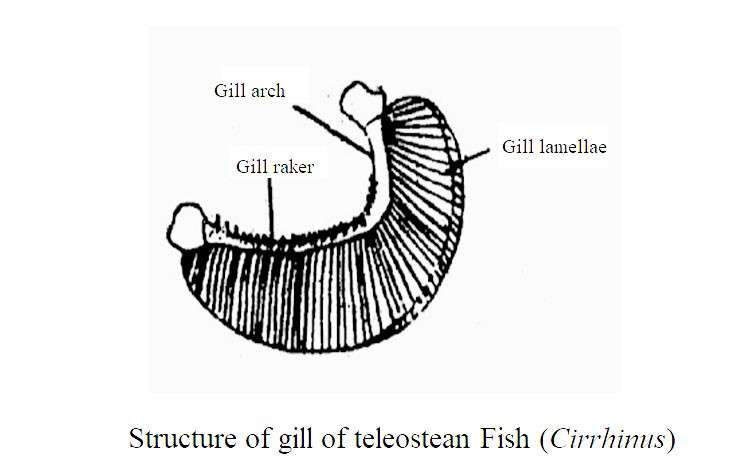
The teleost usually has four pairs of gills. Each gill has a large lower wing and a smaller upper wing which are mainly composed of seratobranchial and epibranchial. An ideal gill of a fish consists of the following parts:
(1) Gill Raker: Gill raker develops at the inner edge of the gill arch. This is the modification of the dermal denticle. It is arranged in two rows. Depending on the diet and eating habits, it can be soft, thin thread-like or hard, flat and triangular or even tooth-like. Each raker is externally covered by an epithelial lining with taste buds and mucus-secreting cells. These taste buds help the fish to understand the chemical nature of the water flowing through the gill opening. The gill rakers are more developed in fish that eat small creatures.
The gill-filament is likely to be damaged or ineffective as small organisms enter through the enternal gill opening during swimming. Typically, gill rakers form a sieve-like structure through which water flows over the gills and filters out the water and protects the brittle gill filaments from solid particles. Different fish show differences in structure of this gill raker.
In plankton-eating fish such as Tenualosa ilisha, Gadusia, Goniolosa, Notopterus, Hypophthalmicthyes, the gill rakers combine to form a filter. Many strainers carry the second and third stage branches of the primary gill rakar and form a dense net. In pike fish (Esox), the gill raker is reduced to form small bony structure which prevent large particles from entering. In closely related species, the formation and numerical differences of the gill rakars are observed.
The mature Alosa alosa has about 80 gill rakar at the bottom of the arch and the Alosa fallax has 30 gill rakar. In Cetorhinus (Basking shark) and Rhinocodon (whale shark), the length of gill rakars are 10-12 cm. In whales, these gill rakars are flattened and functionally formed into baleen plates. Other sharks do not usually have gill rakar. It has been reduced in crossopterygians.
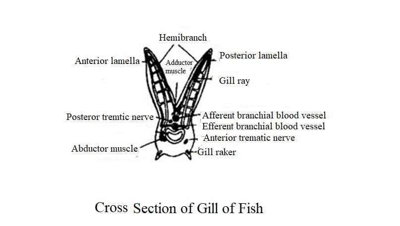
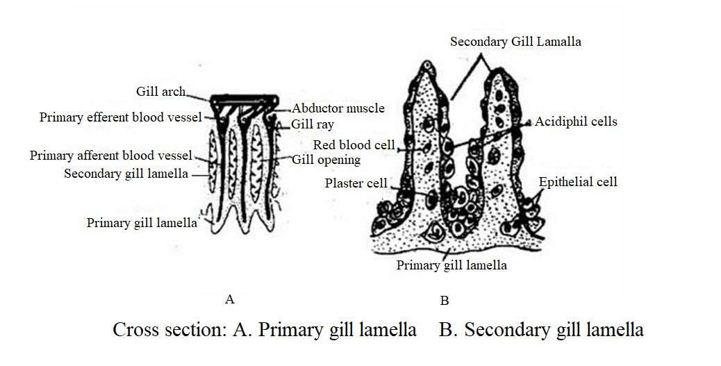
(2) Gill Arch: Each gill arch is surrounded by an efferent and afferent branchial blood vessel and nerve. It is externally surrounded by numerous mucous glands, eosinophilic cells and a thick or thin epithelium with taste buds. Fish living in different ecological habitats vary in number and extent of mucus glands and taste buds. Each gill arch consists of at least one set of abductor and one set of adductor muscles that play a role in the movement of the gill filament (primary lamellae) during respiration.
The abductor muscles attach to the proximal edge of the gill ray from the outside of the gill arch. The adductor muscle resides in the interbranchial septum and enters the opposite gill-ray by crossing each other. There are two known types of adductor muscles and their arrangement. In some teleosts such as Channa striatus, Rita rita, the adductor muscle is generated from the base of the gill-ray on one side and enters the gill-ray on the other side obliquely. In some other species such as Labeo rohita, Tenualosa ilisha, Cyprinus carpio, due to the enlargement of the interbranchial septum, this muscle is generated halfway through the gill-ray and adjacent to the gill-ray on the other side.
(3) Gill filament or primary lamellae: Each gill arch has a two rows of gill filament or primary gill lamellae which are placed outside of the pharyngeal cavity. In most teleosts, the interbranchial septum between the two rows of lamellae is shorter, so the two rows of lamellae are exposed at their distal ends. In Labeo rohita and Tenualosa ilisha, the septum extends to the lower half of the primary lamellae. Due to the diversity of foods, the size, shape and number of primary lamellae vary in different fish.
Species that live actively and rely entirely on aquatic respiration (such as Catla catla, Cirrhinus mrigala, Wallago attu, Mystus seengala) have numerous long gill filaments. However, fish that live inactive or have aerial respiratory organs that have few and and reduced filaments. The gill lamellae consist of two hemibranch that alternate with each other (e.g. Labeo rohita) or they may be attached to the gill arch (Channa striatus).
Usually the gill lamellae in each row are independent of each other. In Labeo rohita, however, the lamellae are consolidated from the base to the tip, and they have narrow crevice-shaped openings. In some river fish, the primary gill lamellae re-divide and form additional lamellae. The lamellae can split in the middle or from the base to form two branches.
Sometimes three to four branches combine to form numerous branches which can form a flower-like structure at one end. The gill ray gives a strong structure to the primary gill lamellae. The gills are composed of partial bones and partial cartilage which are connected to the gill arch and are attached to each other by fibrous ligaments. Each gill ray is divided into two branches. Its nearest edge forms a pathway to the efferent branchial blood vessels.
(4) Secondary Lamellae: Each primary lamellae or gill filament has numerous secondary lamellae on either side. These lamellae have a flattened, leaf-like structure that serves as the main field of gas exchange. Depending on the species living in different ecological habitats, their shape, size and density depend on the unit length of the gill filament. Secondary lamellae are usually free from each other but may merge at the distal end of the primary lamellae.
There are 10-40 secondary lamellae on each side of the primary lamellae. They are numerous in active species. Each secondary lamellae has a central blood vessels rich organs consisting of columnar cells and is surrounded by a basal membrane and the external epithelium. In some fish, this lamellar epithelium is smooth, but other fish it is uneven and has microridge and microvilli.
The epithelium on the surface of the secondary lamellae has grooves and ridges that contribute to increase the respiratory level. One of the structural features of the teleost gill is the presence of columnar cells that are separated by an epithelial covering on the other side of a lamella. Each cell has a central part with an extended part at each end. These enlarged parts are called piller cell flanges that overlap the surrounding cells and form a wall of blood vessels in the lamella. The pillars of the base membrane material connect the secondary lamellae horizontally to the base membrane on the other side and give a structural firmness to the secondary lamellae.
Studies have shown that columnar cells in each row of Channa form a complete divider between blood vessels. Such a system helps in the formation of a narrow water flow and can create reverse flow of blood and water at the microcirculatory level. These columnar cells prevent the contraction and expansion of the blood-rich space and regulate the type of blood flow through the secondary lamellae.
(5) Gill area: The relative number and size of gill lamellae determine the respiration area of the fish’s gills. The overall gill area of a fish species is determined by the overall length of the primary lamella, the number of secondary lamellae, and the average bilateral lamella region. The overall respiration area varies depending on the fish habitat. Generally faster swimming fish have more gill regions and per mm than inactive species. The gill filament contains numerous gill lamellae. Half of the gill area of aerial respiratory fish is directly proportional to the gill permeability efficiency.
During the physical growth of the fish, as the body weight increases, the filtration efficiency of the gill increases. The efficiency of the gill as a respiratory organ also depends on the distance of diffusion. For this reason, obstruction of gas exchange occurs between blood and water. This obstruction is made up of lamellar epithelium, basal membranes, and columnar cell flanges, and it is less common in aquatic respiratory fish than in aerial respiratory fish.
Aquatic respiratory fish have higher diffusion efficiency due to larger gill regions and shorter diffusion distances. However, aerial respiratory fish have a shorter gill area and a wider diffusion distance. According to Saxena (1958), the gill area is much smaller in aerial respiratory fish such as Heteropneustes and Clarias than in aquatic respiratory fish.
Table-9.1: Comparative gill region / area of different fish
|
Species Name |
Number of gill lamellae per mm of gill filament |
Gill region (sqmm Gm-body weight) |
|---|---|---|
|
Scomber scomber |
31 |
1158 |
|
Mugil cephalus |
27 |
954 |
|
Centropristes striatus |
21 |
458 |
|
Opsanus tau |
11 |
197 |
|
Heteropneustes fossilois |
21 |
359 |
|
Clarias batrachus |
19 |
295 |
|
Channa striatus |
15 |
318 |
(6) Gill Epithelium and Branchial Glands: The epithelium surrounded by lamellae is usually two-layered, but in Anabas and Clarias, electromyographs showed a multi-layered epithelium with 5-16 mm thick. The epithelium of these fish usually contains amoebocytes and lymphocytes. Five types of specialized cells are found in fish gills, viz
(1) Ideal goblet type mucus glands– Mucus glands are oval or pear shaped and are ideal goblet type. They form a thin covering through the secretion of acids and relaxing glyco-proteins, thus the respiratory epithelium is protected from dehydration.
(2) Acidophil granular cells;
(3) Basophil mast cells- which act as reservoir of heparin.
(4) Bi or tri-nucleated glandular cells and
(5) Chloride Cells – The presence of chloride cells in the interlamellar epithelium of the filament is thought to be common and which controls the movement of chloride ions through the gill epithelium. Among the above cells, mucus glands and chloride cells are notable.
Fate of Interbranchial septal:
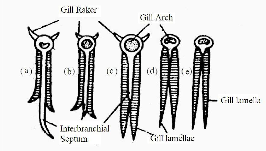
In primitive fish it consists of thick and hard fibrous tissue. The gill arch is made up of many flexible cartilages. The gill arch is located on the inside of the septum. Each arch is semicircular. The lower part of the gill arch is attached to the inside of the gill-filament. Degeneration of interbranchial septum occurs in the course of evolution. In cartilaginous fish, the interbranchial septum is larger than the row of gill-filaments. It covers the gill opening as fold in the back. In Chimera, rows of the gill-filament are slightly shorter in length than the interbranchial septa.
Fig.: Fate of Gill arch and Interbranchial Septum: Elasmobranch (b) Holocephali (c) Holostei (d and (e) Teleosts
In primitive bony fish such as sturgeons and Acipencer, the septa become smaller and extend to the midline of the row. The same condition is seen in Labeo rohita and Tenualosa ilisha, but in other fish this septa becomes shorter. In Salmon, Rita rita and Channa striatus, interbranchial septa looks like as intermediate condition of cartilaginous and bony fish. The gill-filaments on both sides of their septa are independent each other. However, rows of gill-filaments adjacent to the septal tip have merged and a narrow opening is seen between the two rows of gill-filaments.
Fate of Spiracle
There is a crevice-shaped opening between the mandibular and the Hyoidian arch, from which the spiracle emerges. Spiracles exist in sharks. The spiracular gill is located at the tip of the spiracular opening with a combinations of few gill-filament. However, in most fish it consists of a rete merabile called pseudobranch or a network of blood vessels. Spiracles are more developed in sketes and rays and have flexible valves. The external gill pores are located on the ventral side.
When resting on the sandy bottom, sand particles enter the gill pouch through the respiratory water stream, so the respiratory current does not enter through the gill opening at this time to stop the entry of external particles, but enters through the spiracle and exits through the gill opening. In the case of larvae in Holocephali, this opening is present but in adult stage, it is closed. In this case, there is no evidence of the presence of pseudobrancgh in the spiracle opening. The shark has a very small number of spiracles in its mature state.
Spiracles are missing in most cases of bony fish. Although the spiracle sac develops. Amia and Lepidosiren do not have a spiracle, but the spiracular sac is greatly reduced. Polypterus has a wide spiracle. It has a single cellular ridge that separates the spiracular sac and the hyobranchial cavity. Spiracle is missing in Crosopterigii. In Latimaria, the spiracle sac is very deep but in the lung fish this sac is much reduced.
Vuscular Supply of a Teleost Gill
The gills contain well-developed blood vessels. Blood flow and water flow cross each other from opposite directions. Such an arrangement ensures efficient exchange of dissolved components between the two liquids. If the direction of flow of two fluid is changed experimentally, the rate of oxygen uptake decreases from 50% to 9%. The folds of the gill-filament help the blood to come in close contact with water for gas exchange.
The gills contain well-developed blood vessels. Blood flow and water flow cross each other from opposite directions. Such an arrangement ensures efficient exchange of dissolved components between the two liquids. If the direction of flow of two fluid is changed experimentally, the rate of oxygen uptake decreases from 50% to 9%. The folds of the gill-filament help the blood to come in close contact with water for gas exchange.
Recent studies have discovered two pathways of blood flow in the gills, namely:
(1) Respiratory pathway and
(2) Non-respiratory pathway or nutritious pathway.
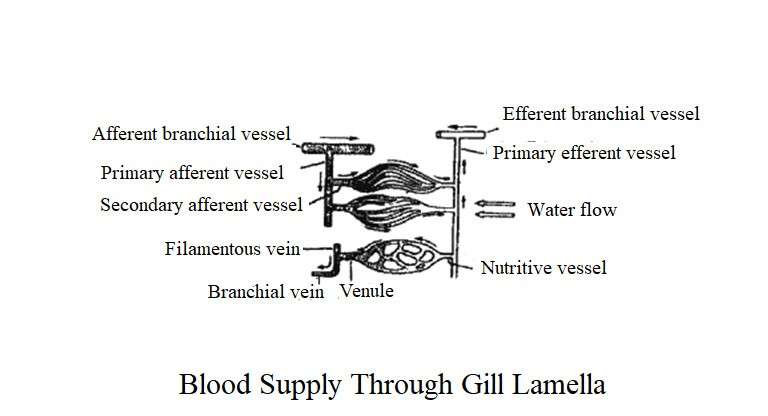
Respiratory pathway is related to respiration. Each afferent branchial blood vessel supplies oxygenated blood from the heart to the gills. This blood vessel is located along the entire length of the gill arch, and numerous primary afferent branches (afferent filamentous arteries) are formed and enter the primary gill lamellae.
Each primary afferent blood vessel splits laterally to produce numerous secondary blood vessels and each secondary gill enters the lamellae. These again produce 2-4 tertiary branches over the gill ray. In the secondary lamellae, these blood vessels break down and intersect with each other by forming lamellae channels. Thus in the secondary lamellae, a blood-rich central core is formed which results in the exchange of gaseous material as blood flows through these channels.
Oxygenated blood reaches the primary efferent blood vessel through the efferent lamellar artery and supplies blood to the edge of the primary gill lamella. At the end, the blood enters the gill arch through the main efferent branchial blood vessel. The non-respiratory pathway is made up of complex structures of the sinuses and veins through which blood passes through the systemic circulation and goes directly to the heart.
It is thought to provide nutrients and oxygen to the filamentous tissue and may also be involved in hormone circulation. This pathway consists of the efferent filamentous artery, the nutritional blood channel, the central venous-sinus, the venules, and the branchial vein.
As a result of the formation of the gill of teleosts, water comes in close contact with the secondary lamellae through which the exchange of gases takes place. Functionally, gills are very efficient and can use about 50-80% of the oxygen present in the water. The gill of the teleost is more efficient than the gill of an elasmobranch due to excessive reduction of the interbranchial septum.
Moreover, due to the arrangement of the efferent and afferent blood vessels, blood flow and respiration occur on opposite sides of the water flow. Such a system is called an inverse flow system where oxygenated water flows from the oral side of the gill to the aboral side and the lamellar blood flows from the aboral side to the oral side, resulting in maximum gas exchange.
Mechanism of Respiration
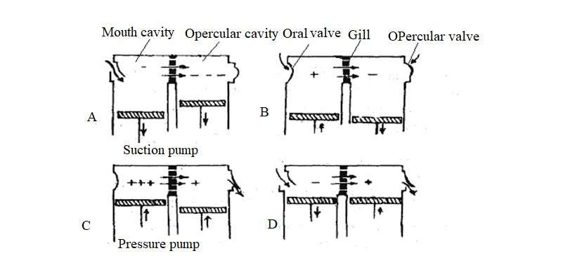
Blood is enriched with oxygen by the inward and outward flow of water through the pharyngeal cavity in the teleost. This process is done by sucking water in the mouth and then coming out through the gill opening. At this time, the rich blood vessels are submerged by the water of lamelle. The pharyngeal cavity plays a role in the release of water by the gills through suction and pressure. Expansion of the lateral wall of the mouth cavity opens the mouth for breathing and enlarges the mouth cavity. The branchistagal ray expands and descends to contract different types of muscles, especially the sternohyoid and palatine elevator muscles.
As the volume of the mouth cavity increases, the water pressure inside it decreases and water enters the mouth cavity due to suction. When the mouth cavity is filled with water, the mouth closes and the volume of the opercular chamber increases as it progresses to the operculum or gill cover, but the opercular cavity closes due to external water pressure. In this way, a low pressure is created in the opercular chamber and water flows over the gills and enters it. Later, their volume decreases due to the exertion pressure of water inside the mouth cavity and the opercular chamber.
Oral valves prevent water from leaking out of the mouth. After the opercula reaches its maximum volume, it quickly moves towards the body. Carbon-dioxide (CO2) rich water escapes through the outer branchial cavity, and the water cannot move in the opposite direction due to the high pressure in the mouth cavity compared to the opercular chamber.
Fig. Showing Dual pumping technique to maintain a constant flow of water through the gills
Respiratory process varies depending on the habitat of the fish. In fast-swimming species, the mouth and opercular opening are more open, so the gills are flooded by an uninterrupted flow of water. The gill chamber of fast-swimming species tend to be smaller than those of inactive species. This technique has also changed in Ostracion, Tetraodon and some mountain river species. Different fish also have different respiratory rates.
Hagfish (Myxiniformes) have two respiratory systems. When the hagfish do not take food, the body without the head is stuck in the mud and in this condition, water enters the gills through the nasopharyngeal cavity. Again, when the mouth is attached to the food, water enters the gill through the egophagocutenius duct and exits. This duct is exposed to the outside through an opening in the back of the last gill pouch.
Physiology of Respiration
Oxygen carrier globular protein, known as hemoglon is placed in the cytoplasm of red blood cells. Hemoglobin is known as respiratory pigment. Each hemoglobin molecule is composed of 5% heme containing iron pigment and 95% globin, polypeptide. Its biggest advantage is that it can absorb different amounts of oxygen depending on the level of tension of gas in the blood. The organism produces energy in the mitochondria through the process of respiration. Oxygen with food is needed to produce this energy. This oxygen is carried from the water to the cells throughout the body by the efferent branchial arteries. Blood plays an important role in the removal of carbon dioxide (CO2) produced by respiration from cells.
Oxygen transport: This is done in two ways, viz
(1) as oxyhemoglobin as a chemical compound through chemical bonding with hemoglobin;
(2) As a physical solution when dissolved in plasma.
(1) As oxyhemoglobin: The hemoglobin molecule consists of four polypeptide chains. Each polypeptide chain contains a globin that is attached to a heme group. An iron molecule (Fe–) is attached to each heme group at the center and an oxygen molecule can be attached to each iron molecule. As a result, one hemoglobin molecule can combine with four oxygen molecules at the same time. Its chemical reaction with oxygen is two-sided and can be expressed by the following equations:
The process of transferring oxygen through hemoglobin is as follows: The process of dissolved oxygen (DO) in water enters the bloodstream through the gills. When tension of oxygen in the blood is less, oxygen is easily combined with hemoglobin. The compound formed by the combination of oxygen and hemoglobin is known as oxyhaemoglobin. Hemoglobin is more attracted to oxygen when the pH of the blood is relatively high.
This relationship of hemoglobin and oxygen with pH is called the Bohr effect. With blood flow, oxyhemoglobin is transported to different parts of the body and oxygen is released in the arteries where the oxygen tension in the arterial blood is low. Free oxygen enters the cell through diffusion. After the oxygen is lost, the oxygen-deprived hemoglobin returns to the gland through intravenous blood. The separation of oxygen from oxyhemoglobin is actually a deoxidation process.
The attachment of oxygen to hemoglobin depends on the amount of oxygen in it, that is, if the amount of oxygen is high, O2 and Hb will combine to form HbO8, and if the amount of O2 is low, HbO8 will break down to separate O2 and Hb. The capillary network around the gill has a high amount of O2 on the side of the efferent branchial artery, so O2 and Hb combine to produce HbO8. The dorsal aorta spreads throughout the body through the efferent branchial blood vessels. Later reaching the cell. Since respiration is continuous, the concentration of oxygen in the cell will always be low, so HbO8 breaks down and O2 is added to enter the cell in the diffusion process.
(2) As a Physical Solution: In this case, without the help of hemoglobin, O2 dissolves in the blood plasma coming directly from the capillary blood vessels to the efferent branchial artery and spreads throughout the body through the dorsal aorta. Eventually it comes in contact with the body cells and releases O2.
Functions of Gills
(1) The main function of gill in fish is respiration. Through the gills, the fish absorb dissolved oxygen (O2) in the water and release the carbon dioxide (CO2) produced by respiration.
(2) In addition to respiration, the gills play a role in maintaining the balance of salt by assisting in the removal of certain wastes.
(3) Freshwater and marine bony fish are excreted in the form of urea and ammonia and take part in excretion. In the case of Cyprinus carpio and Carassius (Gold fish), nitrogenous wastes are excreted 6-10 times more through the gills than in the kidneys.
(4) Gill raker plays an important role in food intake through food selection.


