Giardia duodenalis (Giardia intestinalis/ Giardia lamblia) is a flagellated parasitic microorganism. It is the only pathogenic protozoan found in the duodenum and jejunum of humans. It is the cause of disease, known as giardiasis which is actually infection of the small intestine. It is found throughout the world. Giardiasis is the most common water-borne, parasitic gastrointestinal disease in the USA. In the USA, about 20,000 people are infected by giardiasis each year.
Systematic Position
- Phylum: Sarcomastigophora
- Class: Zoomastigophora
- Order: Diplomonadida
- Family: Hexamitidae
- Genus:Giardia
- Species: Giardia duodenalis
You might also read: Wuchereria bancrofti: Morphology, Life Cycle and Pathogenesis
Morphology
Its life cycle consists of two phages: An active form, called Trophozoite and an inactive form called a cyst.
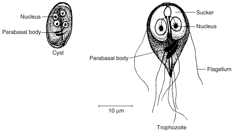
Trophozoite: It is heart-shaped with 10-20 µm in length. It is bilaterally symmetrical having two axostyles, two nuclei with large central karyosome and four pairs of flagella. It looks like a tennis or badminton racket when viewed flat, and like a longitudinally split pear in profile view. The dorsal surface is convex and the ventral surface is concave with a sucking disc. In fresh preparation it has a dancing motion. The trophozoites can attach to the lining of the small intestine using a sucker. It cannot spread the infection to others because it does not live long outside of the body while the cyst can live long time outside the body. When the cyst is ingested, the acid of the stomach stimulates the cyst and produces into disease causing trophozoites.

Cyst: The cyst is oval in shape with 8-14 µm long and 7 µm broad. The contents shrink away a little from the posterior pole. Axostyles lie lengthwise from the anterior pole. Two nuclei in immature and four nuclei in mature cysts are clustered at one end or lie in pairs in two poles. The remains of the flagella may be seen inside the cytoplasm. Generally, the cysts are present in the faces of infected people as a result infection is spread from people to people by contamination of food. It also spreads by direct fecal-oral contamination. Cysts also live in freshwater such as streams and lakes causing water-borne disease, giardiasis.
Life Cycle
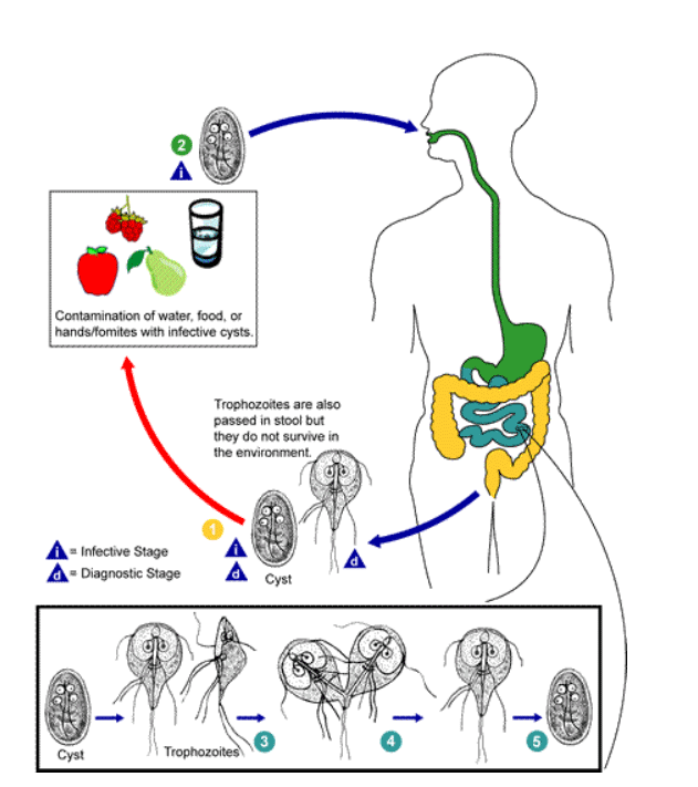
Infection occurs by ingestion of cysts. The cyst excysts in the duodenum into two trophozoites which then multiply by binary fission into large numbers and colonize in the duodenum and upper part of jejunum. Encystment occurs in the colon.

Pathogenesis
Parasites as well as host factors are important. Natural history of infection varies markedly-infection may be aborted, transient, recurrent or chronic. Children are more liable to clinical giardiasis than adults. Trophozoites adhere to epithelium but they do not cause invasive or locally destructive alterations. Loss of brush boarder enzyme activities-may cause lactose intolerance and significant mal-absorption in a minority. The surface of trophozoites is covered by a family of related cysteine-rich proteins that undergo surface antigenic variation and may contribute to prolong and repeated infections.
Clinical Features
It can present as: (1) Asymptomatic-cyst found in stool without any symptom. (2) Acute diarrhea-abdominal pain, bloating, anorexia, nausea, vomiting. (3) Chronic diarrhea-Diarrhea is not prominent, symptoms recurrent and mal-absorption very common. Some patients may become lethargic and flatulent and lose weight.
Common Symptoms of Giardiasis
- Nausea
- Fatigue
- Diarrhea or greasy stools
- Vomiting
- Loss of appetite
- Weight loss
- Bloating and abdominal cramps
- Abdominal pain
- Excessive gas
- Headaches
Laboratory Diagnosis
Stool Examination: Excreted intermittently so examination of 3 or more stool samples on alternate day is advised. Faecal specimen has an offensive odour, pale colored, fatty and float in water.
Microscopic Examination: Trophosoites and or cysts are found in freshly passed stool. Trophozoites are mainly found in mucous. Trophozoites lashes the flagella vigorously making a tumbling sort of movement.
Sting test: Trophozoites may be detected in duodenal aspirates.
ELISA (Enzyme-linked immune-absorbent assay) of stool is a specific and sensitive rapid diagnostic tool.
On investigation there may be mal-absorption of xylose and vitamin B12 and lactose intolerance.
Treatment of Giardiasis
Some specific antibiotics are commonly used to treat giardiasis:
- Antibiotic such as Metronidazole is used to treat giardiasis. In this case, you should take it for five to seven days with single dose.
- Tinidazole is also used to treat giardiasis with a single dose.
- For children, Nitazoxanide is a popular option due to its liquid form. It needs to take only three days to treat giardiasis.
- Besides these, Paromomycin is another antibiotics which needs three doses over the course of 5 to 10 days.
Prevention of Giardiasis
- Maintaining good hygiene;
- Do not drink contaminated water;
- Always drink purified water;
- Do not take contaminated food;
- You have to wash hand before taking food or preparing the food;
- During cleaning the hands, you should use soap;
- Soap should also be used after using toilet;
- During swimming in pools, ponds and lakes, you have to keep the mouth closed;
- Never eat uncooked fruits and vegetables;
- You have to drink packaged drinking water during travelling;
- You should use condom every time during practicing oral-anal sex;
You might also Read: Leishmania donovani-Morphology, Life Cycle, Leishmaniasis and Its Prevention
