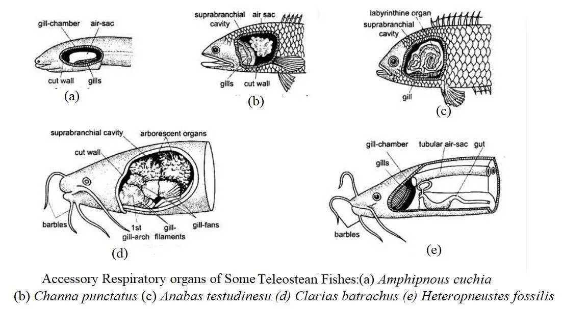
In addition to the gills, all other respiratory organs that exist in different fish are called accessory respiratory organs. For adaptation to special environments, these accessory respiratory organs are developed as an additional part of the gills in fish. In most cases, tropical freshwater fish are seen to develop these organs, but this is very rare in marine fish. In some cases, especially in the summer and when the water level drops, these organs are found in tropical freshwater and mountain river fish to meet the demand for extra oxygen. Some fish have been cut to the ground for a short time to protect themselves from excessive drought. Some fish have such a high metabolic rate that they cannot be met by the oxygen in the water, which has led to the development of some accessory respiratory organs for aquatic or terrestrial respiration.
Accessory respiratory organs of fish can be divided into aerial and aquatic. Of these, aerial respiratory organs are more important for the life of fish. In many fish, it helps in the normal functioning of the gills and ensures extra oxygen.
There are 140 species of aerial respiratory organs in the teleost. When these fish spend a part of their lives on land, they use these organs in case of crisis period. The following table showing the description of some air breathing fish:
Table: Some air breathing fish
|
Name of Family |
Fish Species |
|---|---|
|
Notopteridae |
Chitala chitala, N. notopterus |
|
Cobitidae |
Lepidocephalichthys guntea |
|
Claridae |
Clarias batrachus |
|
Heteropneustidae |
Heteropneustes fossilis |
|
Channidae |
Channa punctatus, C. striatus,C. marulias, C.gachua |
|
Symbranchidae |
Symbranchus bengalensis |
|
Amphipnoidae |
Amphipnous (Monopterus) cuchia |
|
Anabantidae |
Anabas testudineas, Trichogaster fasciatus |
|
Gobidae |
Periophthalmus vulgaris, Boleophthalmus boddaerti, Pseudapocryptes lanceolatus |
|
Mastacembelidae |
Mastacembelus armatus, M. pancalus, M. aculeatus |
|
Amblycipitidae |
Amblyceps mangois |
Various types of accessory respiratory organs have developed in different species of fish. The following is a description of the different types of accessory respiratory organs in fish:
Pharyngeal Diverticula as Respiratory Organ
In snake-headed fish such as Channa punctatus, C. striatus, C. marulius and Cuchia cuchia, the accessory respiratory organs are relatively simple. All these fish can survive out of the water for a short time in an extremely dry environment due to their habit of breathing in the air. The fish of both groups formed a pair of sac-like projections from the gills to exchange gas. In the case of Cuchia, the accessory respiratory organs are relatively simple and consist of a pair of air chambers.
These air chambers develop from the pharynx. The air chambers are further covered by a thin layer of blood vessel-rich epithelium. These air chambers have a simple sac-like structure. These chambers act as a lung-like reservoir. In Channa striatus, the blood vessel-rich epithelium of the air chamber is folded to form some alveoli and the gill filament is reduced in size.
In the case of Amphipnous cuchia, a pair of blood-vessel rich sacs protruding from the pharynx above the gills act as accessory respiratory organs. This projection has been exposed in the 1st gill pouch in the front. Physiologically this projection acts as a lung. The gills are severely reduced and the remaining 2nd and 3rd gill arches have gill filament like some buds. In the 3rd gill arch, there is a fleshy blood vessel-like epithelium. The Periophthalmus has a very small pair of pharyngeal projections which are formed by a layer of blood vessel-rich epithelium.
In the case of these fish, the mouth closes when the pharyngeal muscles contract. The air from the air sac is expelled and goes to the opercular chamber through the exhalent pore. The latter exits through the external branchial chamber. On the other hand, when the pharyngeal muscles are relaxed, the mouth is opened and the air sac is filled with air.
Buccopharyngeal Epithelium as Respiratory Organ
The pharyngeal epithelium of some fish is rich in numerous blood vessels, making it more conductive. It is simple or developing folds that acts as an efficient respiratory organ. It is a tongue-like growth which is formed from the mouth and pharynx. Such respiratory organs are found in fish like Periopthalmus, Boleophthalmous, Amphipnous, Electrophorous etc. It helps absorb oxygen directly from the atmosphere. All of these tropical fish come out of the water and spend most of their time jumping and walking around wet damp places, especially the roots of mangrove forests. It was previously thought that a blood-rich tail helps in respiration. However, modern fisheries scientists do not want to accept it.
An Integument as Respiratory Organ
The skin of Anguilla anguilla, Amphipnous cuchia, Mudskipper (Periophthalmus and Boleopthalmus) is rich in blood vessels and plays a role in aerobic respiration like a frog. Before the development of gills, embryos and larvae of many fish are able to breathe through the skin. The larvae of many fish have numerous blood vessels in the folds of the middle fin that play a role in respiration. The operculum of sturgeon and many catfish is more rich in blood vessels which is used as an accessory respiratory organ. Amphipnous and Mastacembelus live in low oxygen rich closed water bodies and their skin plays little role in respiration. However, it plays an important role in the absorption of gaseous oxygen. When the fish move into or out of the muddy pond, the secretion of skin glands protects the fish from being dry in the aerial environment.
Outgrowth of Pelvic Fin as Respiratory Organ
In the case of lung fish Lepidosiren, they have thin blood-rich out growths of the pelvic fins which play a role in respiration. During reproduction, males develop this out growth that acts as a complement to the respiratory system at a particular time.
Swimbladder Act as Lung
The air sac is an important bouyant organ, but in some fish it acts as a lung. In the case of Amia and Lepisosteus, the membranes of the air sac become enriched and form a lung-like structure. The air sac of Polypterus resembles the superficial lung and a pair of pulmonary arteries originating from the last pair of epibranchial arteries have dilated into it. In terms of the structure and function of the air sac of Diponoids, it is exactly like the lungs of tetrapods. In Neoceratodus, it is a single lobed but in Protopterus and Lepidosiren it is a two lobed. In this case, the formation of spongy alveoli inside the lungs increases. Since these fish cannot use gills during aestivation, they breathe through their lungs. The pulmonary arteries, originating from the last pair of epibranchial arteries of the lungs of Dipnoids like Polypterus is expanded in it.
Alimentary Canal as Respiratory Organ
The stomach or intestines of some fish have changed specifically for aerial respiration. After inhalation, air reach the alimentary canal of these fish and are stored for some time in a special part of it. The following changes have taken place in the digestive tract of fish for exchange of air:
- The wall of the stomach and intestines become significantly thinner, and as a result of excessive contraction of the muscles, it becomes virtually transparent.
- The inner layer of the digestive tract is surrounded by a single layer of epithelial cells. There are no mucus-secreting cells or glands, but the epithelium is rich in blood vessels.
- Excessive amounts of circular muscle fibers are reduced and longitudinal muscle fibers form an extremely thin layer.
Such intestinal respiration is found in loach (Misgurus fossilis) and some fish of the family Siluridae and Loricaridae. In addition to the digestive tract, the intestines of fish of the family Cobitidae play an important role in respiration. In some species the digestive tract (intestine) plays a role in digestion and respiration at short intervals. In other fish, the digestive tract does not play a role in respiration in winter, but it does help in respiration in summer. The internal epithelium of the intestine mainly plays an important role in digestion. Cobitis (giant loach of Europe) comes above the water level and takes a certain amount of air and passes it backwards through the stomach and intestines. Misgurus fossilis has a swelling on the back of the stomach that is covered with a thin layer of blood. This swelling plays the role of accessory respiration. After the gas exchange, the gas escapes through the anus. In Callichthyes, Hypostomus, and Doras, the superior blood vessels acts as a respiratory organ through the intake and excretion of water. The intestinal wall of all these fish is transformed and thinned to reduce muscle layer.
Opercular Chamber Modified for Aerial Respiration
In some species the inhaled air passes through the gill pouch and accumulates in the opercular chamber for some time. In the posterior region of the skull, the opercular chambers form two balloon-like structure. Later, their walls become folded and air escapes through small external branchial openings. Gas exchange plays a role in enriching the membranes of the opercular chamber with a thinner and more abundant blood vessel. This condition is seen in Periopthalmus and Boleopthalmus.
The following structural changes are seen in Periophthalmus:
- Opercular bones are thin and elastic.
- The opercular chamber is enlarged and extends from the base of the basibranchial to the top of the gill arch. Air pockets develop in the wall of the respiratory epithelium.
- In the branchiectal apparatus on both sides, a kind of safety valve develops which is controlled by muscles.
- The epithelial blood vessel of the opercular chamber and branchiostegal membrane is enriched.
- Complex techniques have been developed to open or close the Inhalent and Exhalent pores.
Development of Diverticula from the Opercular Chamber
In highly specialized aerial respiratory fish, sac-like projections develop from the dorsal surface of the opercule or gill pouch. These air chambers or opercular lungs are located at the top of the gills and they have a special structure like labyrinth organs which increase the level of respiration. Accesory respiratory organs are present in fish like Heteropneustes, Anabas, Clarias, Trichogaster, Macropodus, Betta etc.. Important modifications of the respiratory organs in some of these fish species are described below:
Heteropneustes fossilis
The accessory respiratory organs of this species are composed of following parts, namely:
- Wing-like expended gills plate;
- Air sac and
- Respiratory membrane.
Like other teleosts, this species has four pairs of gills, but the gill lamellae are reduced. From these gill arches, four pairs of extended gill plate develop. The gill filaments are attached laterally to the inside of the first gill arch to form the first wing. The second wing is well developed and it resembles an umbrella, the outer gill filaments of the third gill arch merge to form the third wing. The filaments of the fourth gill arch merge to form the fourth wing. In addition to all these wings, there is a pair of simple sac-like structures extending from the supra-branchial chamber to the middle of the caudal region at the posterior side. These are elongated tubular structures with thinner, more reticulate walls that are located within the myotome of the body.
Air sac receives blood from the fourth efferent branchial blood vessel on each side through a branch called a thick-walled afferent respiratory tract. It produces numerous lateral blood vessels that carry blood to the respiratory island. The respiratory membrane of the air sac has numerous folds and crevices so that there are areas rich in blood vessels and without blood vessels. The first region contains numerous respiratory islands. The islands in the lamellae are shortened to form air sacs and wings.
Clarias batrachus
In this species, the suprabranchial organs play an important role as accessory respiratory organs. It consists of the following organs, namely:
- Dendritic organ,
- Supra-branchial chamber, and
- Fan-like organ.
Below is a description of their structure and respiration technique:
Dendritic organ: It is a tall tree-like structure that develops from the top edge of the 2nd and 4th gill arches on each side. This dendritic organ consists of numerous marginal swellings. Each swelling has a cartilaginous core covered by a vascular membrane. Each swelling consists of 6 folds, which proves that each such swelling is made up of 8 gill filaments.
Supra-branchial chamber: A pair of supra-branchial chambers which rich in superior blood vessel that has a tree-like structure. The supra-branchial chamber has developed into a diverticulum rich in blood vessels derived from the gill pouch.
Fan-like organs: The entrance to the suprabranchial chamber is covered by a fan-like structure. The gill filaments present in the dorsal wall of the gill arch combine to form this structure. The wall of the suprabranchial organ is covered by a thin outer epithelium layer and contains intracellular space separated by pilaster cells. Blood comes here through the afferent and efferent blood vessels of the gill arch and helps the fish in aerial respiration. This suprabranchial chamber has incoming and outgoing pores. These fish come to the surface of the reservoir and take atmospheric air from the pharyngeal cavity and enter the opercular chamber through the incoming pore located between the 2nd and 3rd gill arches. After the exchange of gas, air enters the operculum chamber through the gill pore located between the 3rd and 4th gill arches. Fan-like structures exist in the 2nd and 3rd gill arches. It helps to absorb air and causes air to be expelled as a result of contraction of the wall of the suprabranchial chamber. Thus the suprabranchial chamber acts as a complement to the lungs.
Anabas testudineus
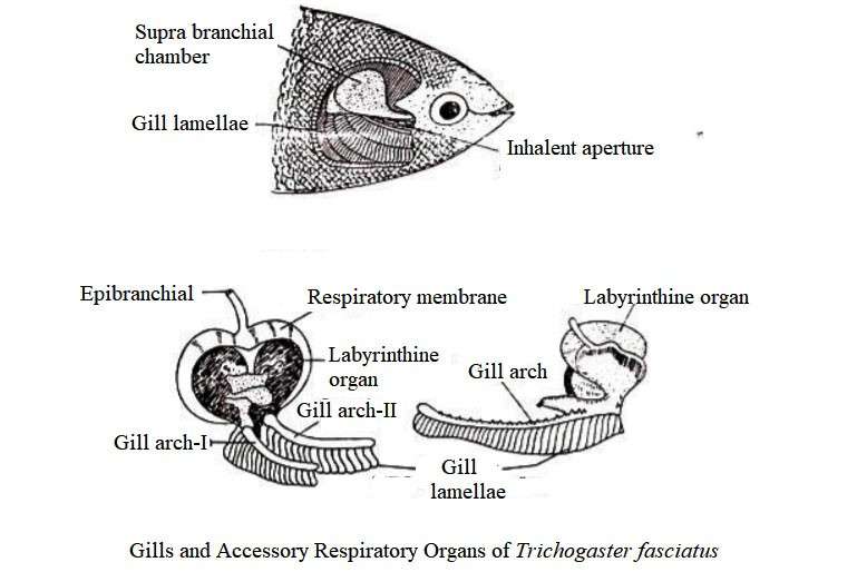
In this species, branchial outgrowth acts as an accessory respiratory organ. In this fish, two broad sac-like protrusions have formed from the dorsal side of the branchial chember. The wall of these epithelium is covered by a layer of epithelium which enriches the blood vessels and enlarges the respiratory area. Each cell has a characteristic rosette-like labyrinthine organ. This organ evolved from the first epibranchial bone and consisted of several concentric plates. The edges of these plates are corrugated and covered by gill-like vascular epithelium.
Each branchial outgrowth maintains free contact with the operculum and bacopharyngeal cavities. The air enters the exterior through the mouth-pharyngeal pores and exits through the pharyngeal pores, and this air is controlled by the entrance valve. This organ can breathe in air through this organ and can travel from one reservoir to another by moving through the operculum and fins. There are sharp spines in the free end of each operculam. During the migration, the operculum expands and grasps the ground with the help of spines and moves forward with the help of thoracic fins and tail. There is a saying that fish can grow on trees. Climbing perch is sometimes seen on the branches of palm trees or other trees which are brought here by kites or crows during terrestrial migration.
Trichogaster fasciatus
These fish have accessory respiratory organs similar to Anabas. It includes the suprabranchial chamber, the labyrinthine organ, and the respiratory membrane. It has stronger labyrinthine organs than the Anabas. Each organ consists of two leaf-like growths. These two growths are composed of loose connective tissue that is covered by a more pigmented blood vessel-rich epithelium.
Function of Accessory Respiratory Organs
These organs act as higher reservoirs of oxygen. Fish with accessory respiratory organs can live comfortably in low oxygenated water. In this case, the fish comes to the surface of the water, fills the air and takes it to the respiratory organs. If these fish are prevented from coming to the surface, they will die of shortness of breath due to lack of oxygen. So the presence of accessory respiratory organs in fish is an adaptive feature. Moreover, the oxygen absorption rate of these organs is higher than the rate of carbon dioxide emitted. Since the gills naturally emit more carbon dioxide. The primary function of the accessory respiratory system is to absorb oxygen.
Origin and Significance of Accessory Respiratory Organs
It is difficult to explain the origin of the fish’s respiratory organs, especially the primary accessory respiratory organs. However, during the embryonic development of the fish, the 5th gill arch does not form the lamellae, but its embryonic gill material initiates the gill arch and these combine to form a gill mass. The aerial respiratory organs develop from this gill mass (Singh 1993). In some species, arches other than the 5th arch take part in the formation of aerial respiratory organs. The development of gill lamellae on the gill arch naturally occurs for aquatic respiration.
According to Singh (1993), the material from which the gills originate in the teleostean fish, air sac or swimbladder develops. The aerial organs of Heteropneustes and Clarias are the modified form of gills (Munshi 1995). These fish have no air sac or completely reduced. Oxygen levels in the atmosphere and reservoirs decrease during the tertiary and quaternary periods of the Cenozoic epoch. Due to the lack of oxygen in rivers and wetlands, the gills are unable to absorb the oxygen required by the body. This is why some teleostian species have developed respiratory organs that help in respiration of gaseous oxygen. Most fish with aerial respiratory organs can live in closed ponds with high oxygen deficiency and ponds with muddy weeds.
There are two opposing theories about the origin of the aerial accessory respiratory organs. The first theory states that short-distance migration from the primary aquatic environment to the terrestrial environment is an inherent feature of some fish. Fish have developed a number of ways to breathe in the air while out of the water. According to the second theory, when the level of oxygen in the water drops significantly, it forces the fish to land. This particular phase of the fish’s life is swallowed by atmospheric air from the land to the respiratory organs. If they are mechanically prevented from coming to the surface in this condition, the fish will die of shortness of breath.
Many bony fish, especially seasonal shallow ponds, have been found to temporarily dry out or the water has become foul-smelling due to the decomposition of aquatic plants. As a result, the habit of breathing in the air for a certain period of time has developed and in addition to fish gills, special types of accessory respiratory organs have been developed. Most of these structures in fish originate from the pharyngeal or branchial chambers. The development of such accessory respiratory organs is essential for adaptation to meet the needs of the respiratory system. In this way the fish can fill the oxygen deficiency in the water which is able to survive in the terrestrial environment for a certain period of time. Accessory respiratory organs develop directly depending on the ability to stay out of the water.
Table: Difference Between Respiratory System of Chondrichthyes and Osteichthyes
|
Chondrichthyes |
Osteichthyes |
|---|---|
|
1. Most cartilaginous fish have 5 gills on each side of the head (except: 4 in Chimaera; 6 in Hexanchus and 6 in Heptranchias which are exposed through holes on the outside). |
1. There are 4 gills on each side of the head. |
|
2. Their gill pores are not covered by operculum (except: Chimaera’s has 4 gills which are exposed through a hole that is covered by operculum). |
2. Their gills are exposed through a hole that is covered by operculum. |
|
3. There is a spiracle between the mandibular and the highway arch in front of the first gill pore. |
3. They have no spiracle. |
|
4. The interbranchial septum between two hemibranch in a holobranch or full gill is larger in size than the two hemibranch. |
4. Such fish have shortened interbranchial septum than hemibranch or in many cases, it is reduced. |
|
5. Their gills (sharks) have one efferent and two efferent blood vessels (except for the holocephalans, which have only one efferent blood vessel). |
5. Each gill arch usually has one afferent and one efferent blood vessel (except for fish such as the lung fish, Labeo rohita, Clarias batrachus, Trichogaster fasciatus, Anabas testudineus, etc.). |
|
6. They usually do not have access to respiratory organs. |
6. In addition to the gills, some of these fish (Anabas, Clarias, Heteropneustes, Channa, Anguilla, Cuchia, Colisa) have accessory respiratory organs. |
|
7. Elasmobranch gills are mostly hemibranch type. Most sharks have a mandibular pseudobranch on each side, a hyoidian hemibranch, and 4, 5, or 6 holobranh (except holocephalans do not have mandibular pseudobranch). |
7. Gills of such fish are mostly holobranch type. Actinopterygian fish have a mandibular hemibranch or pseudobranch on each side, 4 holobranch (exception: Latimeria does not have a mandibular pseudobranch). |


