Echinococcus granulosus is a parasitic worm which belongs to the family Taeniidae under order Cyclophyllidea of class Cestoda. It is also known as Dog tapeworm, Hydatid worm or Hyper-tape worm. It is found all over the world. The larval form causes hydatid disease in man. This disease is also known as human echinococcosis or hydatidosis. At present, it causes major health problems in many countries.
Systematic Position
- Phylum:Platyhelminthes
- Class: Cestoda
- Order: Clyclophyllidea
- Family: Taeniidae
- Genus:Echinococcus
- Species:Echinococcus granulosus
- Definitive Host: Dog, wolf, fox and jackel
- Intermediate Host: Sheep, cattle, pig, human
- Stage found in human: Larval stage.
Morphology of Echinococcus granulosus
Adult Worm
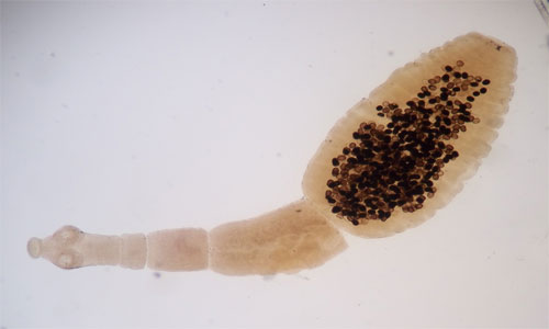
Echinococcus granulosus is a small tapeworm which can grow 3 to 6 mm in length. It comprises of a scolex, neck and 3 or 4 segments. The scolex has 4 suckers and a rostellum with two rows of 30 to 36 hooks. The neck is short and thick.

The anterior first segment is immature, the second segment is mature and third segment or fourth segment (when present) is a gravid segment. Each gravid proglottid contains an average number of 823 eggs. The terminal segment is the longest and broadest.

Eggs
Eggs are spherical in shape and resemble the egg of Taenia solium and Teania saginata. It contains a hexacanth embryo with 3 pairs of hooks. The egg is infective to human, cattle, sheep and other herbivorous animals.
Larval Forms
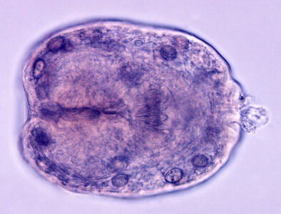
Larval forms are found within the hydatid cyst. It is the scolex of the future adult worm.
Life Cycle of Echinococcus granulosus
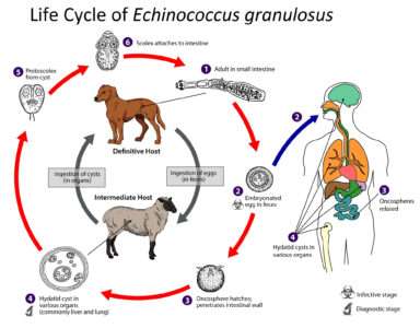
The eggs are discharged with the feces of the definitive hosts such as Dog, wolf, fox and jackel. An itermidiate host such as Sheep, cattle, pig, etc swallow the eggs while grazing in the field and human particularly children swallow the eggs due to intimate handling of infected dogs or other definitive hosts.
In the duodenum, hexacanth embryos are hatched out. About eight hours after infestation, the embryo penetrates through the mucous of the intestine and enters the tributaries of portal vein. It is carried to the liver and may be arrested (liver acts as the first filter). Some embryos may pass the liver, enter the pulmonary circulation and filter out lungs (the second filter). A few embryos may enter the systemic circulation and are trapped in various organs.

Whenever the embryos settles, it forms a hydatid cyst. From the inner surface of the cyst, brood capsules with a number of scolices develop. A definitive host such as dog becomes infected by ingestion of the larva with the infected viscera of an intermediate host such as cattle. The scolex becomes evaginated and develops into an adult worm in 6 to 7 weeks, Thus the cycle is repeated.
Hydatid Cyst
A hydatid cyst may be 1 to 20 cm in diameter. The cyst wall is secreted by the embryo. The cyst wall consists of the following 2 layers:
Outer or circular layer (Ectocyst): It is a laminated hyaline membrane of 1 mm thickness. It is the protective layer.
Inner or germinal layer (endocyst): It is a cellular layer consisting of a protoplasmic mass containing a number of neucei. This vital layer forms (a) brood capsules with scolices(b) daughter cysts (c) hydatid fluid and (d) outer layer.
(a) Brood capsules and scolices: Brood capsules sprout from the germinal layer and consist of only germinal layer. The scolices ( 5 to 20 or more) develop on the inner surface of brood capsules. The fully developed scolices have invaginated sucker and a circle of hooklets. The brood capsules may detach from the cyst wall and then may disintegrate liberating free scolices to form the grains of hydatid sand.
(b) Daughter Cyst: Daughter cysts develop inside the mother cyst. It consists of an outer laminated and inner germinative layer from which brood capsules and scolices develop and also grand-daughter cysts may develop. The daughter cysts mostly grow inwards and are called endogenous daughter cysts. Exogenous cysts may be formed , e.g. in the bone. If the cyst ruptures within the host, the scolices may develop into daughter cysts at different parts of the body.
Hydatid Fluid: It shows the following characteristics-
- Hydatid sand is the sediment of hydatid fluid. It consists of liberated brood capsules, free scolices and loose hooklets.
- If the antigen is released due to rupture or absorption then there may occur anaphylactic symptoms. The antigen is used for immunological tests.
- Clear, colorless with specific gravity of 1005-1010 and pH of 6.7.
Acephalocysts: These are the cysts where brood capsules are not developed or are developed without any scolices.
Pathogenesis
The following symptoms are expressed when hydatid disease/ echinococcosis or hydatidosis occur in man:
- Abdominal pain;
- Nausea;
- Vomiting;
- Chronic cough;
- Chest pain;
- Shortness of breath;
- Anorexia;
- Weight loss;
- Weakness;
- General malaise;
- Hepatic failure, etc.
Diagnosis
- Ultrasonography imaging is the best technique to diagnose of both cystic and alveolar echinococcosis.
- Different serological tests are also effective to detect specific antibodies.
- The best available technique is the ELISA which is suitable for the detection of antigens in feces.
- PCR (polymerase chain reaction) is also employed to detect the parasite from DNA isolated from feces or eggs.
- X-rays, CAT scans etc. can also be done to diagnose the echinococcosis.
Treatment
The treatment for echinococcosis is often expensive and complicated. In many cases, it requires extensive surgery and need prolonged drug therapy. Besides these, the following treatment methods should be applied:
- The most common treatment is the surgical removal of the hydatid cysts.
- Cyst manipulation should be performed. In this case, special caution should be required.
- Percutaneous treatment of the hydatid cysts can be done. In this case, cyst puncture, aspiration of the liquids, Injection, and re-aspiration technique should be applied.
- In very recent days, the new treatment is the Benzimidazole-based chemotherapy.
Prevention
- Anthelminthic vaccinations should be given to dogs and intermediate hosts especially sheep.
- To avoid perpetuating infection, dogs and intermediate host should be kept in separate place.
- Disposal of carcasses and offal should be done properly.
- Livers and lungs containing hydatid cysts should be boiled for 30 minutes.
