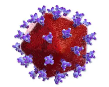
Human Immunodeficiency virus or HIV is the causative agents of acquired immunodeficiency syndrome (AIDS). It attacks the immune system of the body and can lead to AIDS. HIV infects and kills the helper T cells, dendritic cells and macrophages. There is no effective treatment of AIDS but if you take proper medical care, HIV can be controlled. HIV is of two types: HIV-1 and HIV-2. HIV-1 has worldwide distribution; HIV-2 is prevalent in West Africa and is much less virulent.
You might also read: Disinfection and Sterilization
Characteristics of HIV
- HIV is the lentivirus subgroup of retroviruses.
- It is spherical in shape, 100-140 nm in diameter.
- The lipid envelop is derived from the host cell membrane.
- HIV has a bar-shaped core surrounded by a envelope.
- The genome of HIV consists of two identical molecules of single-stranded RNA.
- The envelope contains virus-specific glycoproteins (gp120 and gp41).
- Three enzymes are located within the nucleocapsid of the virion: reverse trancriptage, integrase and protease.
- Reverse transcriptase is the RNA-dependant DNA polymerase. This enzyme transcribes the RNA genome into the proviral DNA.
- Three genes gag, pol, and env are required for replicating retrovirus. In addition, the genome RNA has six regulatory genes.
- Internal ‘core’ protein p24 is encoded by gag gene.
- The pol gene encodes different protein including reverse transcriptase.
- The env gene encodes gp160 that is cleaved to form two envelope glycoproteins, gp120 and gp41.
- HIV has been subdivided into subtypes (clades) on the basis of different base sequence of gp120.

Important Antigens of HIV
- Gp120 and gp41 are the virus specific envelope glycoproteins. The gene that encodes gp120 mutates rapidly, resulting in many antigenic variants.
- P24, the group specific antigen is located in the core. It is an important serological maker of infection.
Replicative Cycle of HIV
1. Attachment, Penetration and Un-coating
The step in the entry of HIV into cell is binding of virion gp120 envelope protein to the CD4 protein on the cell surface. Fruther binding of gp120 with chemokine receptors is required for the entry of HIV. T-cell-tropic strains of HIV bind to chemokine receptor CXCR4 and the macrophase-tropic strains bind to chemokine receptor CCR5.
In the next step, virion gp41 protein mediates fusion of viral envelope with the cell membrane and the virion enters the cell.
2. Gene Expression and Genome Replication
After uncoating, the virion RNA-dependant DNA polymerase transcribes the genome RNA into double stranded DNA which integrates into the host cell DNA. Integration occurs by integrase (virus encoded endonuclease). Immature virion forms in the cytoplasm.
3. Release
Cleavage of the immature virion as buds from the cell membrane by viral protease results in the mature infectious virion.
Transcription
- Sexual Contact: It is the most common amongst male homosexuals. It is also common in bisexual males.
- Intravenous drugs abusers.
- Blood Transfusion of infected blood.
- Administration of infected blood constituents such as factor VIII.
- Vertical transmission: Infected mothers to newborns.
Pathogenesis
HIV infects CD4 helper T cells and kills then resulting in suppression of cell-mediated immunity.
On entry, the dendrite cells that line the mucosa of genital tract are affected. After this the local CD4 helper T cells become infected. HIV binds with CD4 receptors of helper T cells with gp120. There is depletion of helper T lymphocytes. It is the cardinal feature. There is direct cytocidal effect.
The virus is first detected in blood 4-11 days after the infection. The cells recruited into the syncytia die. HIV also kills uninfected helper T cells by indirect mechanisms. The consequences of CD4 T cell dysfunction caused by HIV infection are divesting because CD4 T lymphocytes play a critical role in immune response. HIV probably acts as ‘super antigen’ and thus activates many helper T cells and leads to their demise.
Monocytes and macrophages play a major role in the dissemination and pathogenesis of HIV infection. Certain monocytes and macrophages have CD4 surface molecules. In the brain, the major cell types infected with HIV are the monocytes and macrophages. Monocytes and macrophages serve as major reservoir of HIV. Lymphoid organs play central role in HIV infection.
Coinfections and superinfections with microbes serve as cofactors of AIDS.
Polycional activation of B cells leads to high immunoglobin level circulating immune complexes and autoantibodies. This can cause autoimmune disease such as thrombocytopenia in AIDS patients.
The main immune system is attacked by HIV in the following three mechanisms.
- Viral DNA is integrated into host DNA. This leads to persistent infection.
- High rate of mutation of env gene.
- Down regulation of class I MHC proteins needed for cytotoxic T cells to recognize and kill HIV infected cells.
Clinical Features
The clinical features can be divided into three phages:
Acute Stage
Acute stage usually begins 2-4 weeks after infection. During this time there is viraemia. Viru is widely disseminated throughout the body during this time. Serological tests may be negative. HIV can be transmitted to others during this period. Individuals may present with fever, sore throat, lethargy and generalized lymphanopathy. Maculopapular rash can occur. Acute stage receives spontaneously in about 2 weeks.
Latent Stage
Clinical latency may last for 10 years. After the initial viraemia a ‘viral set point’ occurs. This represents the amount of virus is being produced, i.e. the viral load and may remain set or constant for years. Though the viraemia is low or absent but a large amount of HIV is being produced within lymph nodes. The disease is in latent stage but the virus itself does not enter a latent stage.
A syndrome called AIDS-related complex (ARC) can occur. It is manifested by persistent fever, weight loss and lymphadenopathy. ARC is often progress to AIDS.
Immunodeficiency Stage
The late stage of HIV infection is AIDS. During this stage the CD4 cell count declines below 400/µl and an increase in the frequency and severity of opportunistic infections. Besides other features, development of severe opportunistic infections and unusual neoplasms are characteristics.
The most characteristics manifestations are Pneumocystis pneumonia and Kaposi`s sarcoma. Many neurological symptoms can occur which are caused by HIV infection or due to opportunistic organisms.
Opportunistic Infections in AIDS
Lesions produced which are shown within brackets:
Bacteria: Mycobacterium tuberculosis, Mycobacterium avium-intracellulare, Nocardia asteroids, Salmonella, Streptococcus, Listeria monocytogenes.
Protozoa: Cryptosporidium (enteritis), Isospora belli(enteritis) and Pneumonia or CNS infection
Helminths: Strongyloides stercoralis (Pneumonia, CNS infection or disseminated infection).
Fungi: Candida albicans (mouth, lung, skin, nails, or disseminated). Pneumocystis canini (Pneumonia, disseminated infection), Cyptococcus neoformans (CNS infection), Coccidioides immitis (Disseminated).
Viruses: Cytomegalovirus (Pulmonary, intestinal or CNS infection). Herpes simplex virus (Localized or disseminated), Hepatitis B Virus, Varicella-zoster virus.
Malignant Neoplasm in AIDS
Non-Hodgkin`s lymphoma (e.g. Burkitt`s lymphoma), and Kaposi`s sarcoma are common. Hodgkin`s lymphoma and anogenital cancer may develop.
Diagnosis of AIDS
Serology: Detection of HIV antibodies. Antibodies to viral core protein p24 or envelope glycoproteins gp41, gp120, or gp160 are most commonly detected. Detectable antiviral antibodies appear in all cases within 6 months. Test kits are commercially available for measuring antibodies by enzyme-linked immunoassay (EIA). Sensitivity and specificity of these tests are over 98%.
A positive test in serum sample must be confirmed by a repeat test in blood donors and suspected cases by ELISA. If the repeat ELISA test is positive, a confirmation test like Western blot is performed.
Western Blot Technique (Confirmed test): The antibodies to HIV proteins of specific molecular weights can be detected.
Detection of Viral Nucleic Acid or Antigens
Amplification assays such as RT-PCR (Reverse transcripts polymerase chain reaction) and bDNA( branched DNA) tests are used to detect viral RNA in clinical specimens. P24 antigen can be detected in plasma by EIA.
Virus Isolation
HIV can be cultured from lymphocytes in peripheral blood. Virus isolation techniques are laborious and tissue-consuming and are limited to research studies.
Quantitative Assays for Proviral HIV-1 DNA
Assays for detecting proviral DNA involve the use of PCR to amplify conserved sequences in the HIV-1 gag or pol gene. Used to diagnose infection in neonates.
Assays for Quantification of Plasma HIV-1 RNA
It is used to monitor the course of disease and the response to antiretroviral therapy in patients. For quantifying plasma HIV-1 RNA, different methods are used: (i)RT-PCR (ii) nucleic acid sequence-based amplification (HIV-1 RNA QT).
They are cytocidal for CD4 helper T cells. They are inactivated by household bleach, Lysol, ethanol.
Treatment of AIDS
To prevent emergence of resistance a combination therapy is used. HAART (Highly active antiretroviral therapy)-two neucleoside inhibitors (zidovudine and lamivudine) and a protease inhibitor (indinavir). Other combinations are also used. These are effective in prolonging life, improving quality of the life and reducing viral load. It does not cure the chronic HIV infection, i.e., a latent infection of CD4 positive cell continues indefinitely.
